
Fig. 4
- ID
- ZDB-IMAGE-260501-4
- Publication
- Szenker-Ravi et al., 2024 - CIROZ is dispensable in ancestral vertebrates but essential for left-right patterning in humans
- All Figures
- Figures for Szenker-Ravi et al., 2024
|
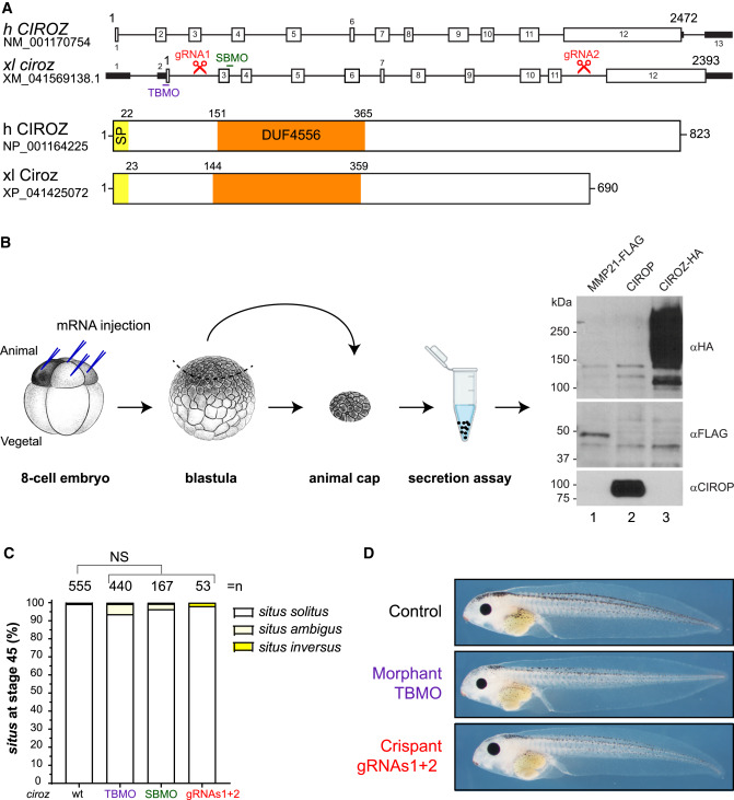
Fig. 4 Xenopus crispants or morphants for ciroz do not exhibit LR defects (A) Depiction of genomic and protein structures of human and Xenopus laevis Ciroz. The Xenopus laevis sequence used is Genbank: XM_041569138.1/XP_041425072. Ciroz protein domains are highlighted: signal peptide (SP; yellow) and DUF44556 (domain of unknown function, orange). The sites targeted by CRISPR gRNA1 and gRNA2 are indicated by two red pairs of scissors in introns 2 and 11, respectively. The sites targeted by the translation-blocking (TBMOs; purple) and splice-blocking (SBMOs; green) morpholinos are also indicated. h, human; xl, Xenopus laevis. (B) Schematic representation of secretion assay using dissociated and cultured animal caps previously microinjected at the 8-cell stage with mRNAs encoding CIROZ-HA, CIROP, or MMP21-FLAG. Western blot performed on the spent supernatant reveals that each of these 3 proteins are readily secreted, consistent with their conserved N-terminal SP. (C) Scoring of internal organs of stage 45 Xenopus larvae. Situs solitus represents the normal situation where the heart is on the left, the gallbladder is on the right, and the intestine rotation is anticlockwise. Situs inversus is the complete mirror image of situs solitus, and situs ambiguus represents any situation in between. n = total number of embryos analyzed from at least 3 independent experiments. Data are mean ± SEM. NS, not significant. Two-way ANOVA with Tukey's test for multiple comparisons (normal condition, situs solitus). wt, wild type. (D) Absence of external phenotype at stage 45. Scale bar, 1 mm.