
Fig. 8
- ID
- ZDB-IMAGE-260428-8
- Antibodies
- Publication
- Attia et al., 2026 - PIKfyve is an essential component of the endolysosomal pathway within photoreceptors and the retinal pigment epithelium
- All Figures
- Figures for Attia et al., 2026
|
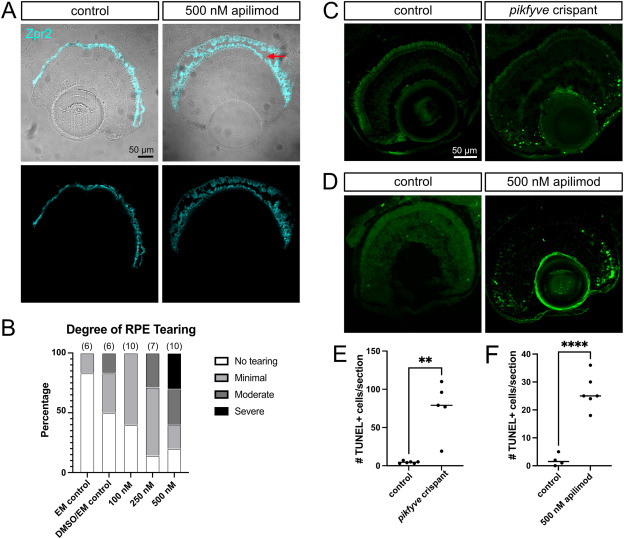
Fig. 8 PIKfyve loss leads to fragility of the RPE and cell death in the retinal periphery. A) Examples of retinal cryosections from 7 dpf larval zebrafish exposed to control solution (1% DMSO in EM) or 500 nM apilimod from 5 to 7 dpf. Zpr2 marks the RPE. Arrow highlights tear in RPE of apilimod-treated fish. B) Qualitative analysis of tearing in RPE according to dose of apilimod from 5 to 7 dpf. Control fish were kept in EM or EM containing 1% DMSO. n = # of eyes. C) Images of TUNEL-labelled retinal cryosections from control and pikfyve crispant fish at 6 dpf. D) Images of TUNEL-labelled retinal cryosections from fish exposed to 1% DMSO in EM control solution or 500 nM apilimod from 5 to 7 dpf. E) Quantification of average # of TUNEL-positive cells per retinal section for pikfyve crispant and control larvae. Each dot represents an individual fish. F) Quantification of average # of TUNEL-positive cells per retinal section for 500 nM apilimod-treated and control larvae. Each dot represents an individual fish. ∗∗p < 0.01, ∗∗∗∗p < 0.0001.
Reprinted from Experimental Eye Research, , Attia, K., Anjum, I., Lingrell, S., Dworkind, C., Benson, M.D., MacDonald, I.M., Hocking, J.C., PIKfyve is an essential component of the endolysosomal pathway within photoreceptors and the retinal pigment epithelium, 110905, Copyright (2026) with permission from Elsevier. Full text @ Exp. Eye. Res.