
Fig. 7
- ID
- ZDB-IMAGE-260428-7
- Antibodies
- Publication
- Attia et al., 2026 - PIKfyve is an essential component of the endolysosomal pathway within photoreceptors and the retinal pigment epithelium
- All Figures
- Figures for Attia et al., 2026
|
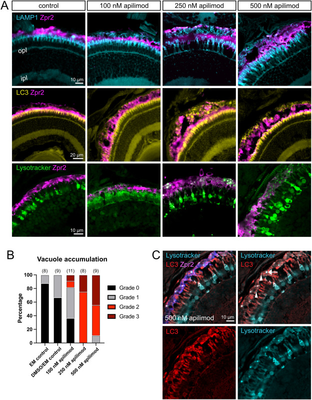
Fig. 7 Characterization of enlarged RPE vacuoles after PIKfyve inhibition. Zebrafish crystal larvae were exposed to a control solution (embryo media (EM) or 1% DMSO in EM), 100 nM apilimod, 250 nM apilimod, or 500 nM apilimod from 5 to 7 dpf. A) Cryosections were labelled with Zpr2 to highlight the RPE and with anti-LC3A/B antibody or anti-LAMP1 antibody. For a third group, the fish were incubated with Lysotracker Red for 2 h prior to fixation. B) Quantification of vacuole accumulation in the RPE. Retinal sections were qualitatively ranked according to number and size of vacuoles in the RPE, from no vacuoles (grade 0) to presence of large vacuoles and/or >25 small vacuoles (grade 3). C) Co-staining of RPE vacuoles with LC3 and Lysotracker reveals that only a subset of vacuoles is labelled with Lysotracker. Arrow indicates vacuole co-labelled with Lysotracker and LC3 and arrows indicate LC3+ vacuoles not labelled by Lysotracker. ipl, inner plexiform layer; opl, outer plexiform layer. (For interpretation of the references to color in this figure legend, the reader is referred to the Web version of this article.)
Reprinted from Experimental Eye Research, , Attia, K., Anjum, I., Lingrell, S., Dworkind, C., Benson, M.D., MacDonald, I.M., Hocking, J.C., PIKfyve is an essential component of the endolysosomal pathway within photoreceptors and the retinal pigment epithelium, 110905, Copyright (2026) with permission from Elsevier. Full text @ Exp. Eye. Res.