
IMAGE
Fig. 6
- ID
- ZDB-IMAGE-260428-6
- Publication
- Attia et al., 2026 - PIKfyve is an essential component of the endolysosomal pathway within photoreceptors and the retinal pigment epithelium
- All Figures
- Figures for Attia et al., 2026
Image
|
Figure Caption
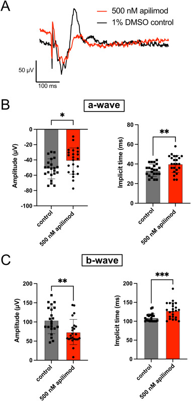
Fig. 6 Decrease in visual function following post-embryogenesis inhibition of PIKfyve. ERG recordings were performed on 6 dpf zebrafish exposed to 1% DMSO in embryo media (controls) or 500 nM apilimod from 4 to 6 dpf. A) Example ERG traces from one control and one apilimod-treated fish. B) Quantification of a-wave amplitude and implicit time. C) Quantification of b-wave amplitude and implicit time. Each dot on the graphs in B-C represents the average measurement of five recordings from an individual fish. n = 22 control fish, 23 apilimod-treated fish. ∗p < 0.05; ∗∗p < 0.01; ∗∗∗p < 0.001.
Figure Data
Acknowledgments
This image is the copyrighted work of the attributed author or publisher, and
ZFIN has permission only to display this image to its users.
Additional permissions should be obtained from the applicable author or publisher of the image.
Reprinted from Experimental Eye Research, , Attia, K., Anjum, I., Lingrell, S., Dworkind, C., Benson, M.D., MacDonald, I.M., Hocking, J.C., PIKfyve is an essential component of the endolysosomal pathway within photoreceptors and the retinal pigment epithelium, 110905, Copyright (2026) with permission from Elsevier. Full text @ Exp. Eye. Res.