
Fig. 5
- ID
- ZDB-IMAGE-260428-5
- Publication
- Attia et al., 2026 - PIKfyve is an essential component of the endolysosomal pathway within photoreceptors and the retinal pigment epithelium
- All Figures
- Figures for Attia et al., 2026
|
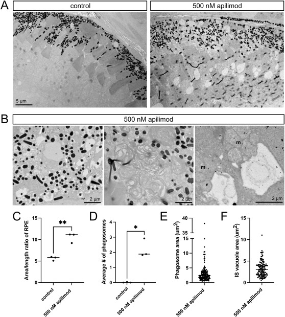
Fig. 5 PIKfyve inhibition post embryogenesis induces outer retina vacuolation and impaired phagocytosis. A) TEM images of outer retina from zebrafish larvae exposed to 1% DMSO in embryo media (controls) or 500 nM apilimod from 4 to 6 dpf. B) Higher magnification TEM images of zebrafish larvae exposed to 500 nM apilimod showing examples of RPE vacuolation (left), a stalled and expanded RPE phagosome (middle), and large vacuoles in two photoreceptor inner segments, just below the mitochondrial clusters (m). C-F) Quantification based on TEM imaging of zebrafish larvae exposed to 1% DMSO in embryo media (control) or 500 nM apilimod from 4 to 6 dpf. C) Quantification of RPE expansion, measured as area/length ratio. Each dot represents the average measurement for an individual fish (n = 3 fish per group). D) Quantification of phagosome number per 10 μm length of RPE. Each dot represents the average measurement for an individual fish (n = 3 fish per group). E) Plot of phagosome area, with each dot representing an individual phagosome. Measurements are pooled from three apilimod-treated fish. F) Plot of inner segment (IS) vacuole area, with each dot representing an individual vacuole. Measurements are pooled from three apilimod-treated fish. ∗p < 0.05; ∗∗p < 0.01.
Reprinted from Experimental Eye Research, , Attia, K., Anjum, I., Lingrell, S., Dworkind, C., Benson, M.D., MacDonald, I.M., Hocking, J.C., PIKfyve is an essential component of the endolysosomal pathway within photoreceptors and the retinal pigment epithelium, 110905, Copyright (2026) with permission from Elsevier. Full text @ Exp. Eye. Res.