
Fig. 4
- ID
- ZDB-IMAGE-260428-4
- Publication
- Attia et al., 2026 - PIKfyve is an essential component of the endolysosomal pathway within photoreceptors and the retinal pigment epithelium
- All Figures
- Figures for Attia et al., 2026
|
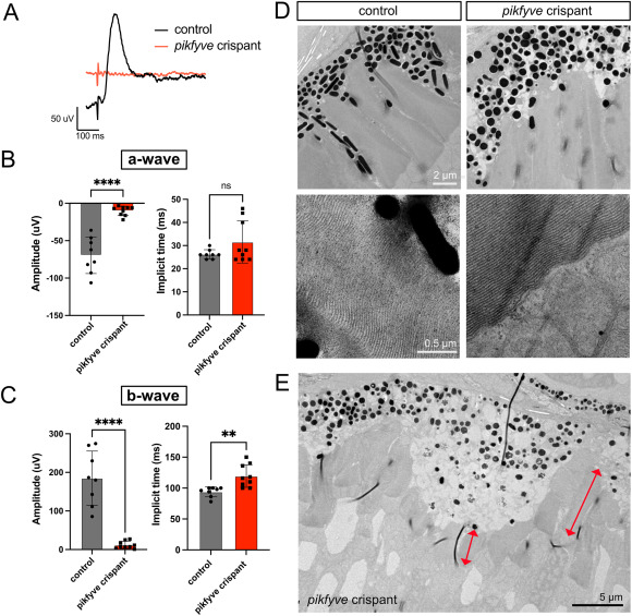
Fig. 4 Absent visual responses of pikfyve crispants despite largely preserved outer segments. A) Representative photopic electroretinography (ERG) traces from pikfyve crispant and uninjected control zebrafish at 5 dpf. B) Quantification of a-wave amplitude and implicit time. Note that the implicit time was difficult to assess in the pikfyve crispants given the lack of a visible a-wave. C) Quantification of b-wave amplitude and implicit time. Each dot on the graphs in B-C represents the average measurement of five recordings from an individual fish (n = 8 uninjected controls, n = 9 pikfyve crispants). F) TEM images of photoreceptor outer segments (OS) in 6 dpf control and pikfyve crispant fish reveals no apparent disorganization of OS discs in crispants. G) In areas of RPE expansion in pikfyve crispants, OS length is reduced (red double arrows indicate OS length for two photoreceptors). (For interpretation of the references to color in this figure legend, the reader is referred to the Web version of this article.)
Reprinted from Experimental Eye Research, , Attia, K., Anjum, I., Lingrell, S., Dworkind, C., Benson, M.D., MacDonald, I.M., Hocking, J.C., PIKfyve is an essential component of the endolysosomal pathway within photoreceptors and the retinal pigment epithelium, 110905, Copyright (2026) with permission from Elsevier. Full text @ Exp. Eye. Res.