
Fig. 2
- ID
- ZDB-IMAGE-260428-2
- Publication
- Attia et al., 2026 - PIKfyve is an essential component of the endolysosomal pathway within photoreceptors and the retinal pigment epithelium
- All Figures
- Figures for Attia et al., 2026
|
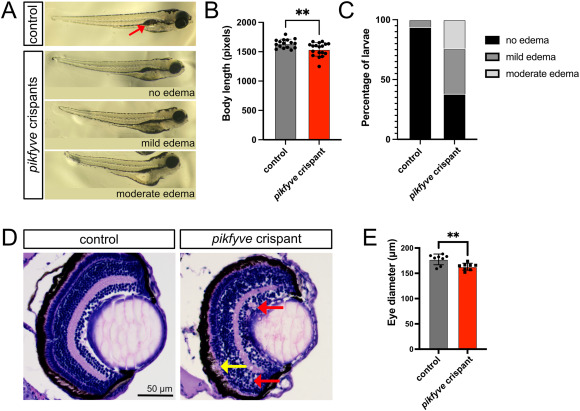
Fig. 2 pikfyve crispant zebrafish larvae exhibit gross morphological changes. A) Brightfield stereomicroscope images of 6 dpf uninjected control and pikfyve crispant embryos, demonstrating that many of the crispant larvae exhibit a shorter body length, edema, and failure to inflate swim bladder (red arrow shows inflated swim bladder in control fish). B-C) Quantification of body length and edema in pikfyve crispants (n = 18 larvae) and uninjected controls (n = 16 larvae). D) H&E stained paraffin sections of eyes from uninjected control and pikfyve crispant 6 dpf zebrafish larvae. Red arrows point to vacuoles in crispant retina and yellow arrow to gap between RPE and outer nuclear layer (ONL). E) Quantification of eye size for uninjected control (n = 9 larvae) and pikfyve crispant fish (n = 8 larvae). ∗∗p < 0.01. (For interpretation of the references to color in this figure legend, the reader is referred to the Web version of this article.)
Reprinted from Experimental Eye Research, , Attia, K., Anjum, I., Lingrell, S., Dworkind, C., Benson, M.D., MacDonald, I.M., Hocking, J.C., PIKfyve is an essential component of the endolysosomal pathway within photoreceptors and the retinal pigment epithelium, 110905, Copyright (2026) with permission from Elsevier. Full text @ Exp. Eye. Res.