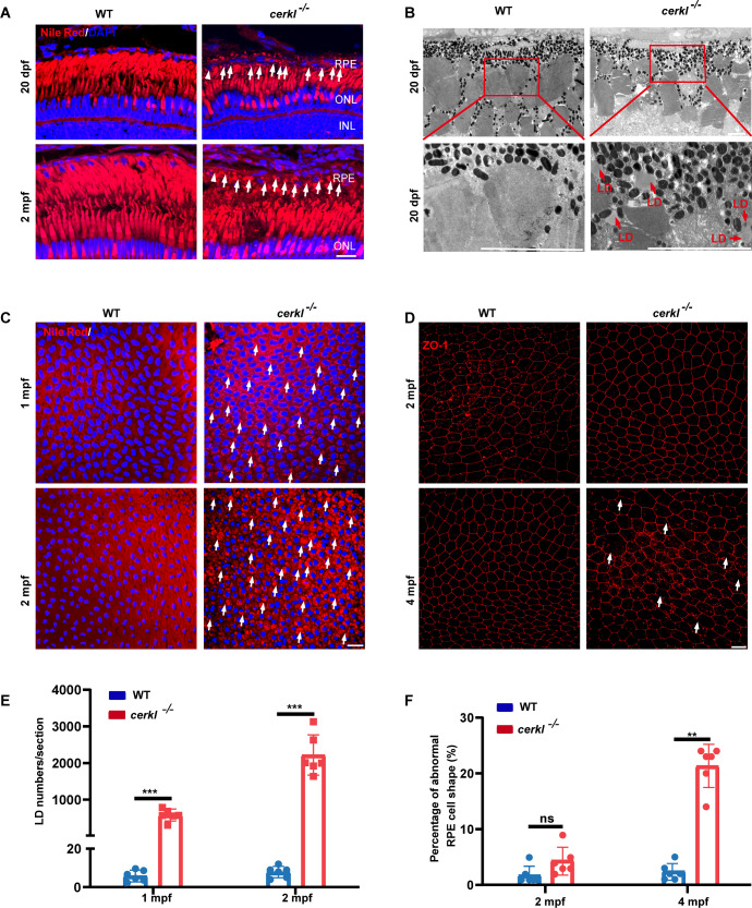
CERKL ablation triggers progressive lipid droplet pathology in RPE. (A) Confocal imaging of LDs in WT and cerkl−/− retina at 20 dpf and 2 mpf zebrafish by Nile Red staining. Blue represents DAPI and red represents Nile Red staining. RPE, retinal pigment epithelium; ONL, outer nuclear layer; INL, inner nuclear layer; Scale bars = 10 µm. (B) TEM analysis of LDs in WT and cerkl−/− zebrafish retina at 20 dpf. LD, lipid droplet. Scale bars = 10 µm. (C) Confocal imaging of LDs in WT and cerkl−/− RPE at 1 mpf and 2 mpf zebrafish by Nile Red staining. Blue represents DAPI and red represents Nile Red staining. Scale bars = 10 µm. (D) Immunofluorescence analysis of RPE cells from WT and cerkl−/− zebrafish at 2 mpf and 4 mpf using anti-ZO-1 antibody. Scale bars = 10 µm. (E) The quantitative analysis of LD numbers shown in C. The results are shown as mean ± SEM. ***, P < 0.001. (F) The numbers of abnormal RPE cells were qualified for each of the images in D. The results are shown as mean ± SEM. **, P < 0.01.
Acknowledgments
This image is the copyrighted work of the attributed author or publisher, and
ZFIN has permission only to display this image to its users.
Additional permissions should be obtained from the applicable author or publisher of the image.
Full text @ Invest. Ophthalmol. Vis. Sci.
Your Input Welcome
Thank you for submitting comments. Your input has been emailed to ZFIN curators who may contact you if
additional information is required.
Oops. Something went wrong. Please try again later.