IMAGE

Figure 2.

ID
ZDB-IMAGE-260423-57
Publication
Hernandez et al., 2026 - Insulin Induces Outer Blood-Retinal Barrier Disruption via Downregulation of Claudin-19
All Figures
Figures for Hernandez et al., 2026
Image
Figure Caption

Figure 2.

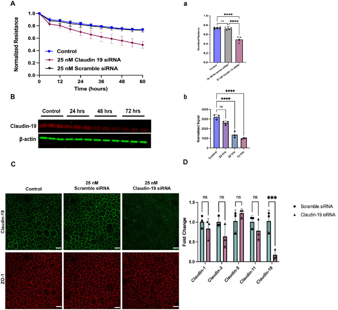
Claudin-19 plays an essential role in maintaining the outer BRB. (A) Real-time barrier-resistant measurement (2000 Hz) of primary porcine RPE cells treated with 25-nM scrambled siRNA or claudin-19 siRNA or were left untransfected. Only final points were quantified. Data are mean ± SD (n = 5). ****P < 0.0001 (ordinary one-way ANOVA with Tukey's test). (B) Western blot analysis of porcine RPE treated for 24, 48, and 72 hours with 25-nM scrambled siRNA or claudin-19 siRNA. β-Actin was used as a loading control. Data are mean ± SD (n = 5). ****P < 0.0001 (ordinary one-way ANOVA with Tukey's test). (C) Immunofluorescence staining of claudin-19 (green) and ZO-1 (red) in porcine RPE cells. Scale bar: 25 µm. (D) mRNA expression of claudin-1, claudin-3, claudin-5, claudin-11, and claudin-19 after 72-hour treatment with 25-nM scrambled siRNA or claudin-19 siRNA. Data are mean ± SD (n = 3). ***P < 0.0001 (two-way ANOVA with Tukey's test); ns, not significant.

Acknowledgments
This image is the copyrighted work of the attributed author or publisher, and ZFIN has permission only to display this image to its users. Additional permissions should be obtained from the applicable author or publisher of the image. Full text @ Invest. Ophthalmol. Vis. Sci.