IMAGE

Figure 5

ID
ZDB-IMAGE-260416-54
Source
Figures for Anderson et al., 2026
Image
Figure Caption

Figure 5

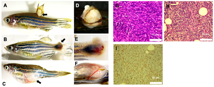
Characterization of ES-like tumors in adult zebrafish. (A) Lateral view of 15-month-old transgenic fish with tumor on dorsal fin. (B) Lateral view of 10-month-old transgenic fish with mass on caudal fin. (C) Lateral view of 12-month-old transgenic fish with mass near anal fin. Black arrows indicate ES-like tumors in (AC). (D) Magnified image of dorsal fin tumor, dorsal view. (E) Magnified image of caudal fin tumor, lateral view. (F) Magnified image of anal fin tumor, ventral view. (G) Histological analysis of the zebrafish anal fin tumor with H&E staining. (H) PAS staining of the zebrafish anal fin. (I) Histological analysis of the zebrafish anal fin tumor upon CD99 staining, which was performed in two independent fish tumors. These findings indicate that a subset of adult Tg(col2a1a:EWSR1::FLI1:pA) zebrafish develop tumors with histological and immunophenotypic features consistent with ES neoplasms, including small round blue cell morphology on H&E staining, glycogen accumulation detected by PAS staining, and expression of the diagnostic cell-surface marker CD99.

Acknowledgments
This image is the copyrighted work of the attributed author or publisher, and ZFIN has permission only to display this image to its users. Additional permissions should be obtained from the applicable author or publisher of the image. Full text @ Int. J. Mol. Sci.