
Image
|
Figure Caption
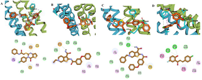
Figure 4.
(A) 3D and 2D binding interactions of F0579-0616, (B) 3D and 2D binding interactions of F0526-1309, (C) 3D and 2D binding interactions of F0526-1306, and (D) 3D and 2D binding interactions of F1190-0509 within the ATP synthase binding pocket. In the 3D view, hits are shown in orange, amino acids in cyan, and the protein backbone in blue and green for chains 6 and 7, respectively.
Acknowledgments
This image is the copyrighted work of the attributed author or publisher, and
ZFIN has permission only to display this image to its users.
Additional permissions should be obtained from the applicable author or publisher of the image.
Full text @ Bioinform. Biol. Insights