
Image
|
Figure Caption
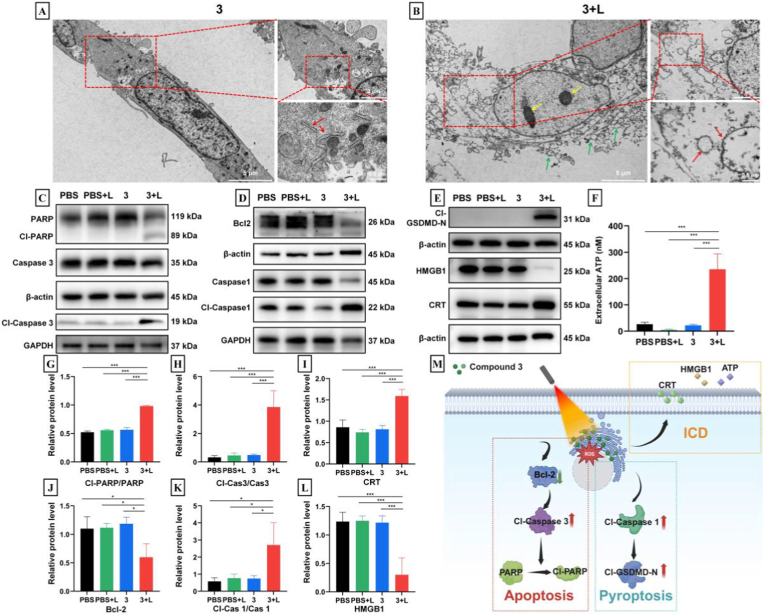
Fig. 6
(A-B) Electron micrographs of 4T1 cells after different treatments. (C-E) Western blotting analysis of apoptosis, pyroptosis and immunogenic death after various treatments. (F) The adenosine triphosphate (ATP) content in the supernatant after various treatments (n = 3). (G-L) Schematic illustration of the molecular mechanism of killing cells under light irradiation (n = 3).
Acknowledgments
This image is the copyrighted work of the attributed author or publisher, and
ZFIN has permission only to display this image to its users.
Additional permissions should be obtained from the applicable author or publisher of the image.
Full text @ Mater Today Bio