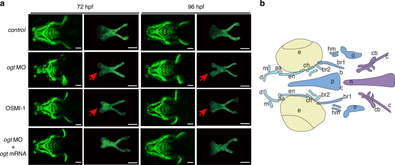
Cleft palate and impaired parasphenoid bone formation caused by the loss of O-GlcNAcylation in zebrafish. a Representative fluorescence ventral views of Tg(sox10: eGFP) transgenic zebrafish larvae. Palatal skeletons were dissected and flat-mounted. The anterior part is facing upwards. The white solid line represents the ethmoid palate. Scale bars, 100 μm. b Schematic diagram of the ventral (up) view of bone structures of 9 dpf zebrafish larvae. aa anguloarticular, br1 branchiostegal ray1, en ectopterygoid, m maxilla, n notochord, o opercle, p parasphenoid, br2 branchiostegal ray2, c cleithrum, cb ceratobranchial 5, ch ceratohyal, d dentary, hm hyomandibular, v vertebrae, bop basioccipital articulatory process
Acknowledgments
This image is the copyrighted work of the attributed author or publisher, and
ZFIN has permission only to display this image to its users.
Additional permissions should be obtained from the applicable author or publisher of the image.
Full text @ Int J Oral Sci
Your Input Welcome
Thank you for submitting comments. Your input has been emailed to ZFIN curators who may contact you if
additional information is required.
Oops. Something went wrong. Please try again later.