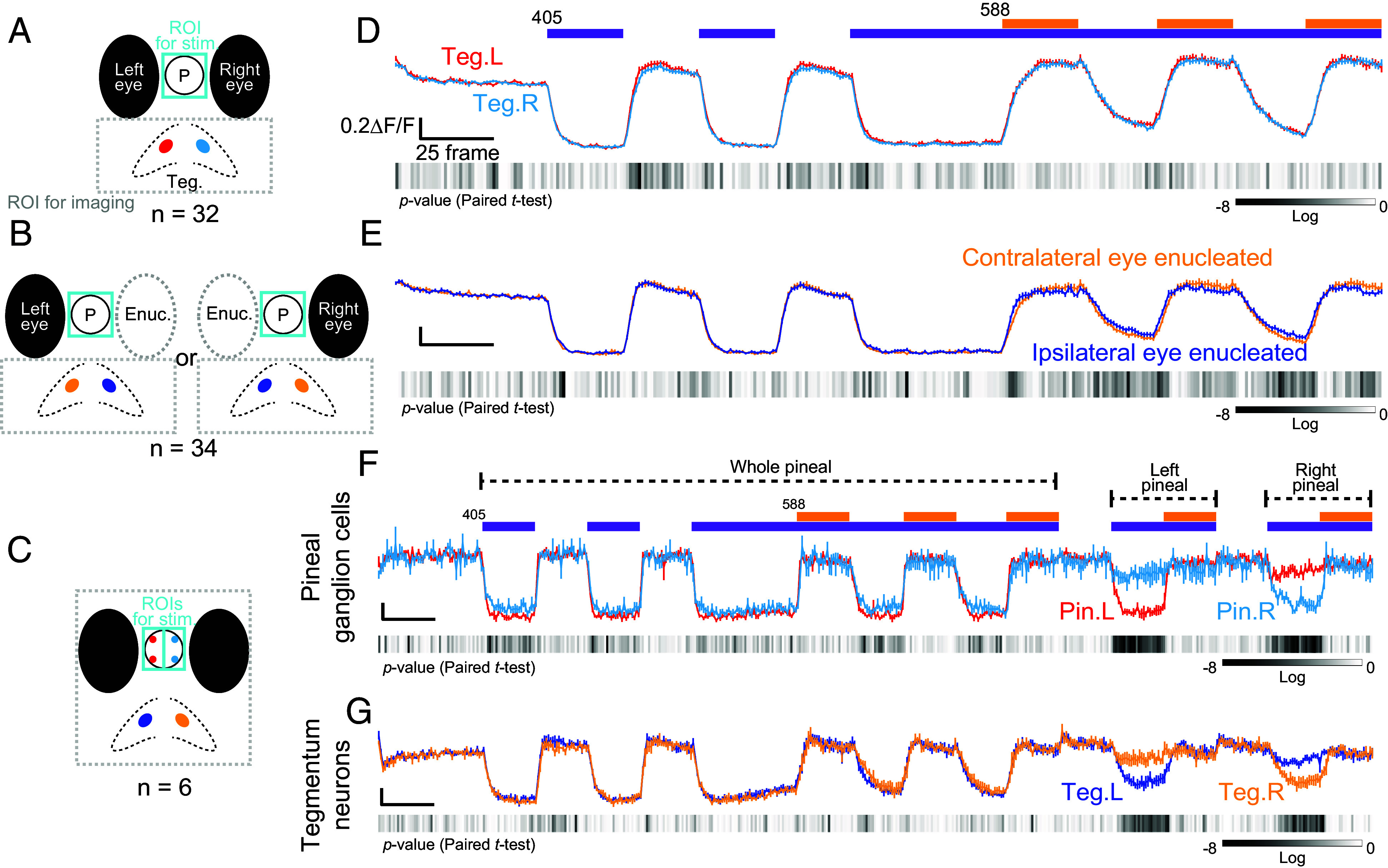
Relationship between photosensory organs and tegmentum in innervation for C-type neurons. (A–C) Schematic representations of the targeted neurons to examine how they are innervated by the eyes and the pineal organ. P, pineal organ; Teg, tegmentum; Enuc, enucleated eye. The C-type neurons in the colored areas were comparatively analyzed in (D–G). (D and E) Calcium change profiles recorded in tegmentum imaging. Colored profiles represent data obtained from left (red) and right (cyan) C-type tegmentum neurons (D) and C-type tegmentum neurons in contralateral (yellow) and ipsilateral (blue) eye-enucleated fish (E). (F and G) Calcium change profiles recorded in whole-brain imaging from limited planes (refer to 11-30 planes in Fig. 3 G, ii and iii), with additional stimuli applied to the left and right pineal organ. The profiles show calcium changes in the left (red) and right (cyan) pineal ganglion cells (F), and in the left (blue) and right (yellow) tegmentum neurons (G). Experiments were performed using 5 to 6 dpf zebrafish larvae.
Acknowledgments
This image is the copyrighted work of the attributed author or publisher, and
ZFIN has permission only to display this image to its users.
Additional permissions should be obtained from the applicable author or publisher of the image.
Full text @ Proc. Natl. Acad. Sci. USA
Your Input Welcome
Thank you for submitting comments. Your input has been emailed to ZFIN curators who may contact you if
additional information is required.
Oops. Something went wrong. Please try again later.