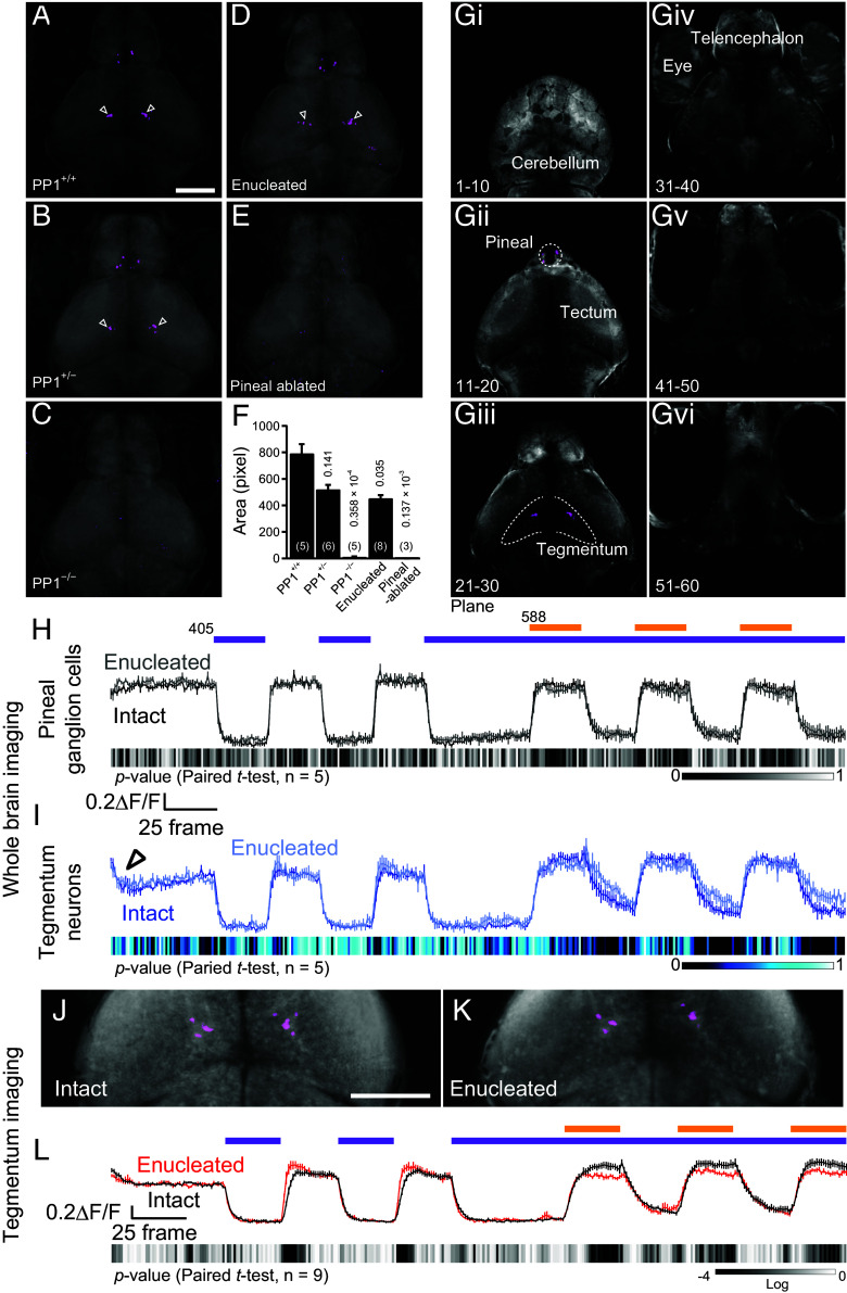
The pineal C-type property based on PP1 is transmitted to tegmentum neurons. (A–E) C-type neuron-visualized images in different genotypes: PP1+/+ (A), PP1+/− (B), and PP1−/− (C), eye-enucleated fish (D), and pineal-ablated fish (E) in whole-brain imaging. (Scale bar: 100 μm.) (F) Quantification of the C-type area in the tegmentum (Dunnett’s multiple comparison test). (Gi–Gvi) Six separate optical brain sections showing that C-type pineal ganglion cells (Gii) and C-type tegmentum neurons (Giii) are present in different planes and that C-type cells were not found in other planes (Gi and Giv–vi). (H and I) Comparison of calcium change profiles in C-type pineal ganglion cells (H) and tegmentum neurons (I) before (intact, black and blue) and after (enucleated, gray and light blue) surgical enucleation (n = 5) in whole-brain imaging. The open arrowhead in (I) shows the decrease in calcium levels upon the onset of two-photon excitation laser in the tegmentum neurons. (J, K, and L) Comparison of C-type-visualized images (J and K) and calcium change profiles (L) before and after surgical enucleation (n = 9) in tegmentum imaging. Data are normalized to the average of frames 26-50 (25 frames before the initial UV exposure in H, I, and L). (Scale bar: 100 μm.) Experiments were performed using 5 to 6 dpf zebrafish larvae.
Acknowledgments
This image is the copyrighted work of the attributed author or publisher, and
ZFIN has permission only to display this image to its users.
Additional permissions should be obtained from the applicable author or publisher of the image.
Full text @ Proc. Natl. Acad. Sci. USA
Your Input Welcome
Thank you for submitting comments. Your input has been emailed to ZFIN curators who may contact you if
additional information is required.
Oops. Something went wrong. Please try again later.