
Image
|
Figure Caption
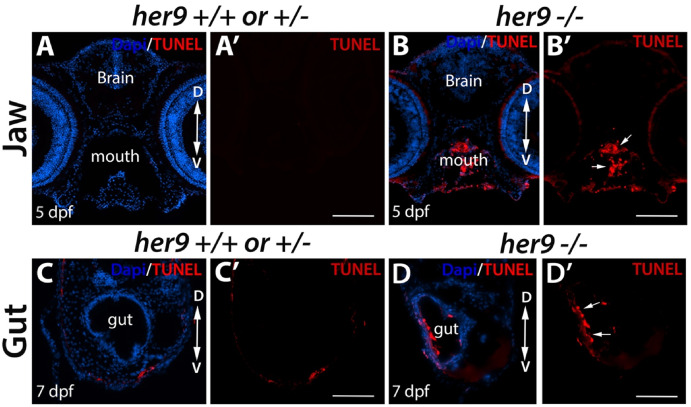
Fig. 8 Loss of Her9 results in apoptosis. A-B′) TUNEL staining of sections through the heads of 5 dpf WT/heterozygous and her9 mutant larvae. TUNEL + cells are detectable in the mouth and jaw region of her9 mutants (arrows). C-D′) TUNEL staining of sections through the gut of 7 dpf WT/heterozygous and her9 mutant larvae. TUNEL + cells are detectable in the gut of her9 mutants (arrows). Scale bars A-B' = 20 μm; C-D' = 50 μm.
Acknowledgments
This image is the copyrighted work of the attributed author or publisher, and
ZFIN has permission only to display this image to its users.
Additional permissions should be obtained from the applicable author or publisher of the image.
Full text @ Differentiation