
|
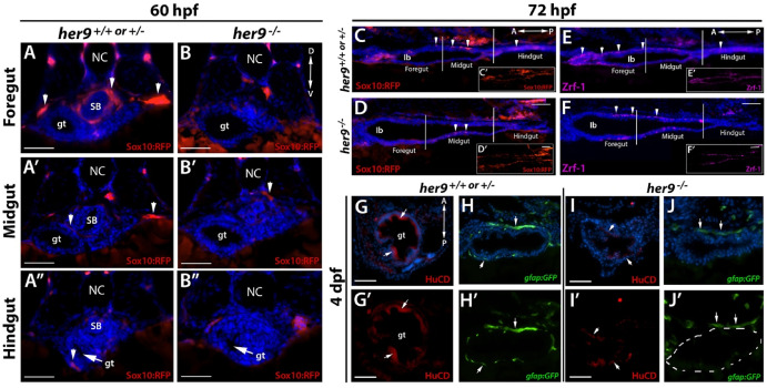
Fig. 5 Loss of Her9 impairs NCC migration and differentiation in the gut. A-B″) Transverse sections through foregut, midgut, and hindgut of sox10:RFP WT/heterozygous and her9 mutant embryos at 60 hpf. While some RFP + cells have colonized all three regions of the WT/heterozygous gut (arrowheads), no RFP + cells are visible in any region of the her9 mutant gut at this stage. C-D) Lateral sections through the gastrointestinal tract of sox10:RFP WT/heterozygous and her9 mutant larvae at 72 hpf. RFP + cells are present in all three regions of the WT/heterozygous gut, but are only detected up to the midgut of her9 mutants. E-F) Lateral sections through the gastrointestinal tract of WT/heterozygous and her9 mutant larvae at 72 hpf immunolabeled for the glial marker Zrf-1. Zrf-1+ cells are absent from the her9 mutant intestinal bulb (Ib) and hindgut. G-J′) Transverse sections through the gut of gfap:GFP WT/heterozygous and her9 mutant larvae at 4 dpf and immunolabeled for the neuronal marker HuC/D. The her9 mutants display fewer enteric neurons and fewer enteric glial cells than their WT/heterozygous siblings. SB, swim bladder; gt, gut; Ib, intestinal bulb; NC, notochord. Scale bars A-B″, G-J' = 50 μm; C-F = 25 μm.