
|
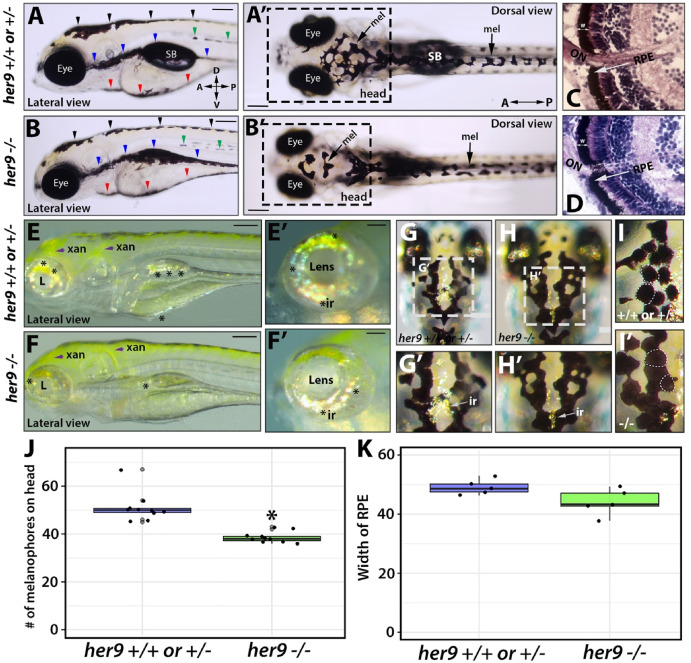
Fig. 3 Her9 mutants display pigmentation defects. A-B′) Lateral (A-B) and dorsal (A′-B′) views of melanophore patterning in WT/heterozygous and her9 mutant larvae at 6 dpf. Black arrowheads, dorsal stripe; green arrowheads, lateral stripe; blue arrowheads, ventral stripe; red arrowheads, yolk stripe; SB, swim bladder. C-D) H&E staining of WT/heterozygous and her9 mutant retina at 7 dpf; the retinal pigmented epithelium (RPE) is indicated with a white arrow. E-F′) Xanothophore (xan) and iridophore (ir) patterning in WT/heterozygous and her9 mutant larvae. Her9 mutants have reduced abundance of iridiphores (asterisks) and greater spread of xanthophores (yellow) than WT or heterozygous siblings. G-I′) Dorsal view of iridiphores and melanophores on the heads of WT/heterozygous and her9 mutant larvae. J) Quantification of melanophores in WT/heterozygous and her9 mutant larvae K) quantification of RPE width WT/heterozygous and her9 mutants. Scale bars A-B′,E, F, G, and H = 20 μm; C-D = 100 μm; E′, F′, G′, H′ and I’ = 40 μm. (For interpretation of the references to colour in this figure legend, the reader is referred to the Web version of this article.)