
|
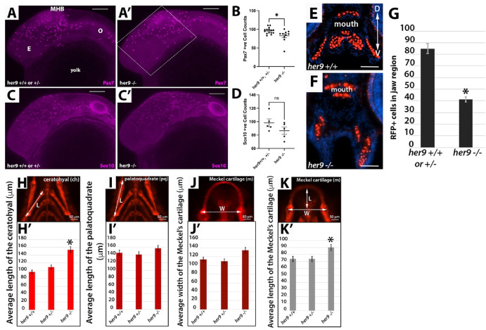
Fig. 2 Loss of Her9 results in reduced CNCC and viserocranium structural defects. A-D) Whole mount immunohistochemistry for Pax7 (A-B) and Sox10 (C-D) at 24 hpf. The box in A′ indicates the region anterior to the midbrain-hindbrain boundary (MHB) where cells were counted for the graphs in B and D. The number of Pax7+ and Sox10+ cells in this region was reduced in her9 mutants relative to WT and heterozygous siblings. E-G) Sox:RFP + cells are reduced in her9 mutant craniofacial region at 5 dpf (n = 8 mutants, n = 13 WT or heterozygous larvae, ∗p < 0.001). H-K′) Analysis of craniofacial structures in WT, heterozygous, and her9 mutant larvae at 5 dpf. Images in H-K are representative WT samples shown for reference. ∗p < 0.05. Scale bars for A-C′, 100 μm; E-F = 50 μm.