IMAGE

Fig. 4.

ID
ZDB-IMAGE-260316-109
Publication
Harvey et al., 2026 - A glial cell derived pathway directs regenerating optic nerve axons toward the optic chiasm
All Figures
Figures for Harvey et al., 2026
Image
Figure Caption

Fig. 4.

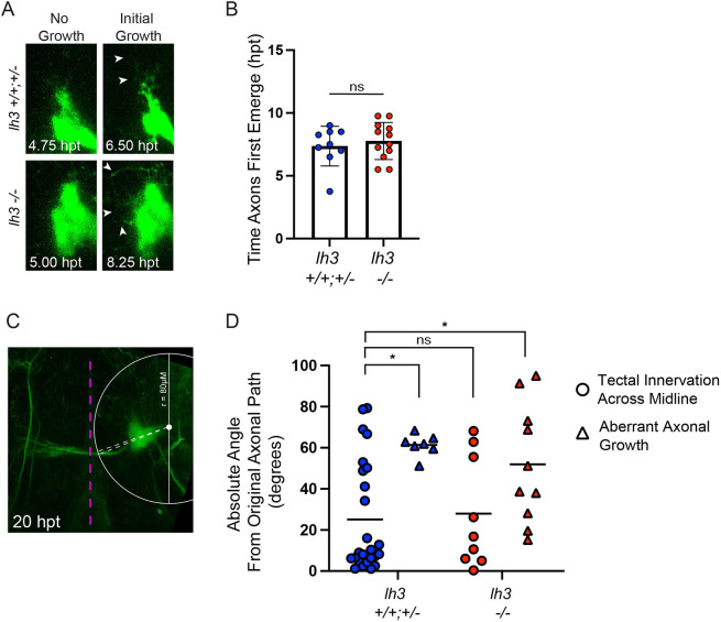
lh3 is required to direct regrowing RGC axons along their original path. (A) Representative frames from live timelapses of lh3 sibling (top) and lh3 mutant (bottom) zebrafish larvae expressing Tg(isl2b:GFP) to label RGCs showing the initial axonal growth that emerges from the injured proximal stump of axons. hpt, hours post-transection. (B) Comparison of the time when axon fascicles first emerge from the proximal stump in lh3 siblings and mutants. Time was determined by the first frame when axon fascicles were observed extending from the proximal stump that extended for at least one other consecutive frame. Data are represented as mean±s.d.; n.s., P>0.05 (unpaired t-test). (C) Representative image of a regenerating nerve in a lh3 sibling at 20 hpt and the mark-up to measure the angle of growing fascicles from the injured stump of axons. The dashed magenta line marks the parasphenoid cartilage. See Materials and Methods for more details on quantifications. (D) Quantification at 20 hpt of the absolute angle of axon fascicles from the end of a projection to the path of the original axonal projection (Tectal Innervation Across Midline: sibling, n=6 nerves; mutants, n=3 nerves; Aberrant Axonal Growth: sibling, n=3 nerves; mutants, n=6 nerves). Individual dots are measured fascicle absolute angles, lines represent means. n.s., P>0.05; *P<0.05 (one-way ANOVA). See Table S1 for individual angles.

Acknowledgments
This image is the copyrighted work of the attributed author or publisher, and ZFIN has permission only to display this image to its users. Additional permissions should be obtained from the applicable author or publisher of the image. Full text @ Development