IMAGE

Fig. 2.

ID
ZDB-IMAGE-260311-64
Publication
Tarbashevich et al., 2026 - Cell-autonomous control coupled with tissue context regulates the cessation of migration at the site of organ development
All Figures
Figures for Tarbashevich et al., 2026
Image
Figure Caption

Fig. 2.

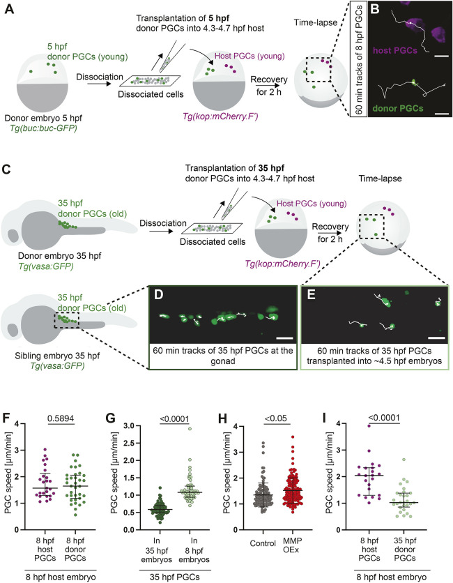
PGC migration speed is controlled by environmental cues and a cell-intrinsic program. (A) Embryos at 5 hpf were dissociated, and the mixture of labeled PGCs (green) and somatic cells (gray) was transferred into wild-type embryos. (B) Tracks of ‘young’ donor PGCs and ‘young’ host PGCs within ‘young’ host (7-9 hpf). Scale bars: 20 µm. (C) Gonad regions of 35 hpf embryos were dissociated. This mixture of labeled PGCs (green) and somatic cells (gray) was transferred into wild-type embryos. (D,E) Tracks of ‘old’ PGCs in control embryos (no transplantation) at 35 hpf (D) and of ‘old’ donor PGCs transplanted into ‘young’ host (E). Scale bars: 30 µm. (F) Speed of ‘young’ host and donor cells within ‘young’ hosts (see B). 12 embryos with 25 PGCs (host); 20 embryos with 35 PGCs (donor). (G) Speed of 35 hpf (‘old’) PGCs at the gonad of control sibling embryos (‘old’) (see D) and within ‘young’ host embryos (see E). 19 embryos and 75 PGCs (35 hpf gonad), 14 embryos and 63 PGCs (35 hpf transplanted). (H) Speed of PGCs overexpressing MMPs, relative to control at 24-26 hpf. Control: 36 embryos, 110 PGCs; MMP overexpression: 36 embryos, 131 cells. (I) Speed of ‘young’ host (8 hpf) and ‘old’ donor (35 hpf) PGCs in ‘young’ host embryos (see C and E). 12 embryos, 22 PGCs (host); 28 PGCs (donor). Unpaired two-tailed t-test. Data are median±error bars (IQR) in F,G,I. In H, data are mean±s.d.

Acknowledgments
This image is the copyrighted work of the attributed author or publisher, and ZFIN has permission only to display this image to its users. Additional permissions should be obtained from the applicable author or publisher of the image. Full text @ Development