
Image
|
Figure Caption
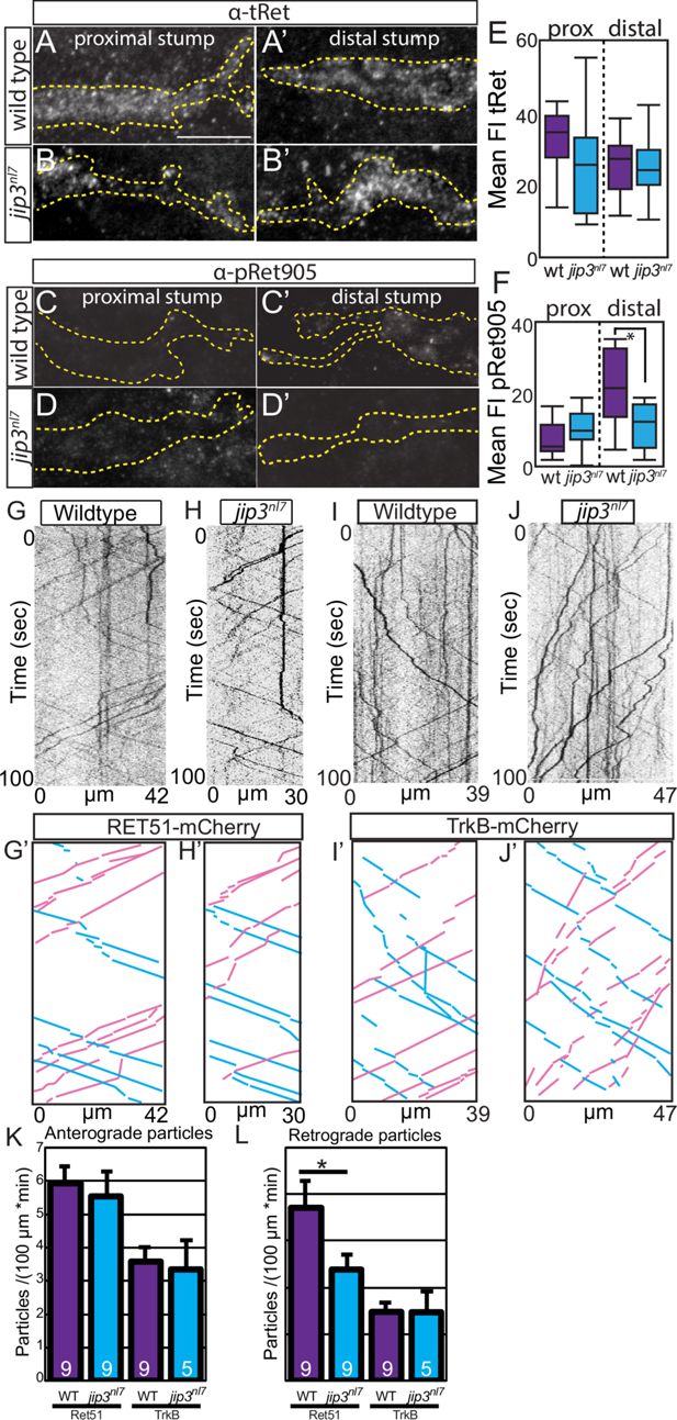
Fig. 5 In Figure 5, panel 5C was inadvertently duplicated in place of panel 5D. These images contain two channels: GFP, which outlines the nerve stump, and pRet905 immunolabeling. The GFP channel (not shown in Figure 5) was used to generate the dotted nerve outline for pRet905 quantification. While the pRet905 image in panel 5D was incorrect due to duplication, the GFP channel and thus the nerve outline was correct.
Acknowledgments
This image is the copyrighted work of the attributed author or publisher, and
ZFIN has permission only to display this image to its users.
Additional permissions should be obtained from the applicable author or publisher of the image.
Full text @ Elife