IMAGE

Fig 5

ID
ZDB-IMAGE-260118-139
Publication
Flannery et al., 2026 - Impact of maternal compensation on developmental phenotypes in a zebrafish model of severe congenital muscular dystrophy
All Figures
Figures for Flannery et al., 2026
Image
Figure Caption

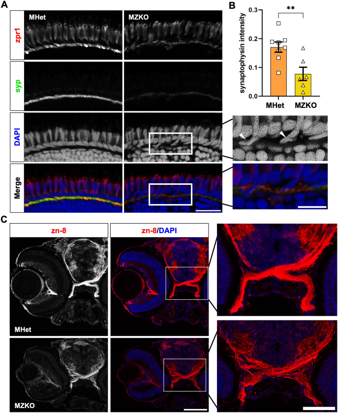
Fig 5 Retinal synapse formation and axon guidance deficits in MZKOs.

A: Fluorescent staining of the outer layers of the retina showing comparable zpr1 (Arr3a) staining in the photoreceptor outer segments and pedicles in the outer plexiform layer, but reduced and disrupted synaptophysin (syp) staining, protrusion of photoreceptor cell bodies in the outer plexiform layer (arrowheads), and disorganization of horizontal cells lining the top of the outer nuclear layer (asterisks) suggesting defects to the ribbon synapses (40X magnification, scale bars: 20 µm for main panels, 10 µm insets). B: Quantification of synaptophysin intensity in the outer plexiform layer showing a significant reduction in MZKOs (**p = 0.0069). C: Fluorescent staining of transverse cryosections with zn-8 and DAPI showing an overall disruption in morphology of the retina with evidence of retinal ganglion cell axon defasciculation in the optic nerves at the chiasm (10X magnification, scale bars: 100 µm for main panels, 20 µm for chiasm insets).

Figure Data
Acknowledgments
This image is the copyrighted work of the attributed author or publisher, and ZFIN has permission only to display this image to its users. Additional permissions should be obtained from the applicable author or publisher of the image. Full text @ PLoS Genet.