
|
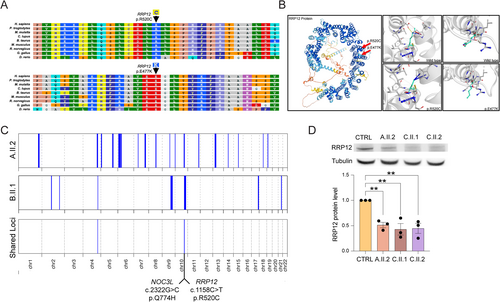
Fig. 2 Genetic and molecular characterization of RRP12 gene variants. (A) RRP12 p.(R520C) and p.(E477K) variants are highly conserved across species. (B) In silico analyses (DynaMut and ColabFold) predict the destabilization of mutated RRP12 proteins. Wild-type and mutant residues are colored in light green and are also represented as sticks alongside the surrounding residues, which are involved in any type of molecular interaction. (C) Homozygosity mapping of probands A.II.2 and B.II.1 showing shared loci of homozygosity on chromosome 4 (hg19: Chr4:155,525,695-159,727,192) and chromosome 10 (Chr10:95,360,964-101,150,256), which includes the RRP12 c.1558C>T and NOC3L c.2322G>C variants. (D) Western blot analysis of RRP12. Quantification of RRP12 revealed a statistically significant decrease in RRP12 in all affected samples compared to the control (P = 0.0029). *P ≤ 0.05; **P ≤ 0.01; ***P ≤ 0.001.