
|
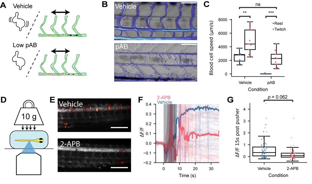
Fig. 3 Blood vessel deformation directly induces Ca2+ events in ECs. (A) Schematic showing effect of 30 μM para-amino blebbistatin (pAB) on cardiac contractility, blood flow, and body motion. (B) Brightfield transmitted light movies were used to quantify blood flow. Composite image showing average image (grey) and speckle amplitude (color), caused by flowing blood cells. Treatment with pAB stopped the heartbeat (Video S2) and largely suppressed cardiac-driven blood flow. (C) Dorsal aorta blood cell velocity in vehicle (0.3% DMSO) and pAB treatment. (A – C) n = 9 fish vehicle, n = 9 fish pAB. Statistical test: Mann-Whitney-Wilcoxon two-sided test. Twitch vs. rest (vehicle): p = 6.2e-3, U = 9.0; Twitch vs rest (pAB): p = 4.1e-4, U = 0; rest (vehicle) vs twitch (pAB): p = 0.66, U = 46.0. (D) Schematic of weight press experiment. (E) Example ΔF/F at 15 s after press for Tg(kdrl:Gal4;vglut2a:Gal4;UAS:GCaMP6s) fish treated with vehicle (0.3% DMSO) or 20 μM 2-aminoethoxydiphenyl borate (2-APB). (F) Fluorescence response vs. time relative to weight push. Solid lines are median over individual vessels. Initial response is dominated by motion associated with application of the weight (shaded region). (G) Fluorescence response 15 s after weight application. (F-G) 92 vessels from n = 11 fish vehicle, 93 vessels from n = 13 fish 2-APB. Statistical test: mixed-effect linear model considering the effects of 2-APB and individual fish, p = 0.062, z=−1.9. Additional supporting data in Figure S3.