
|
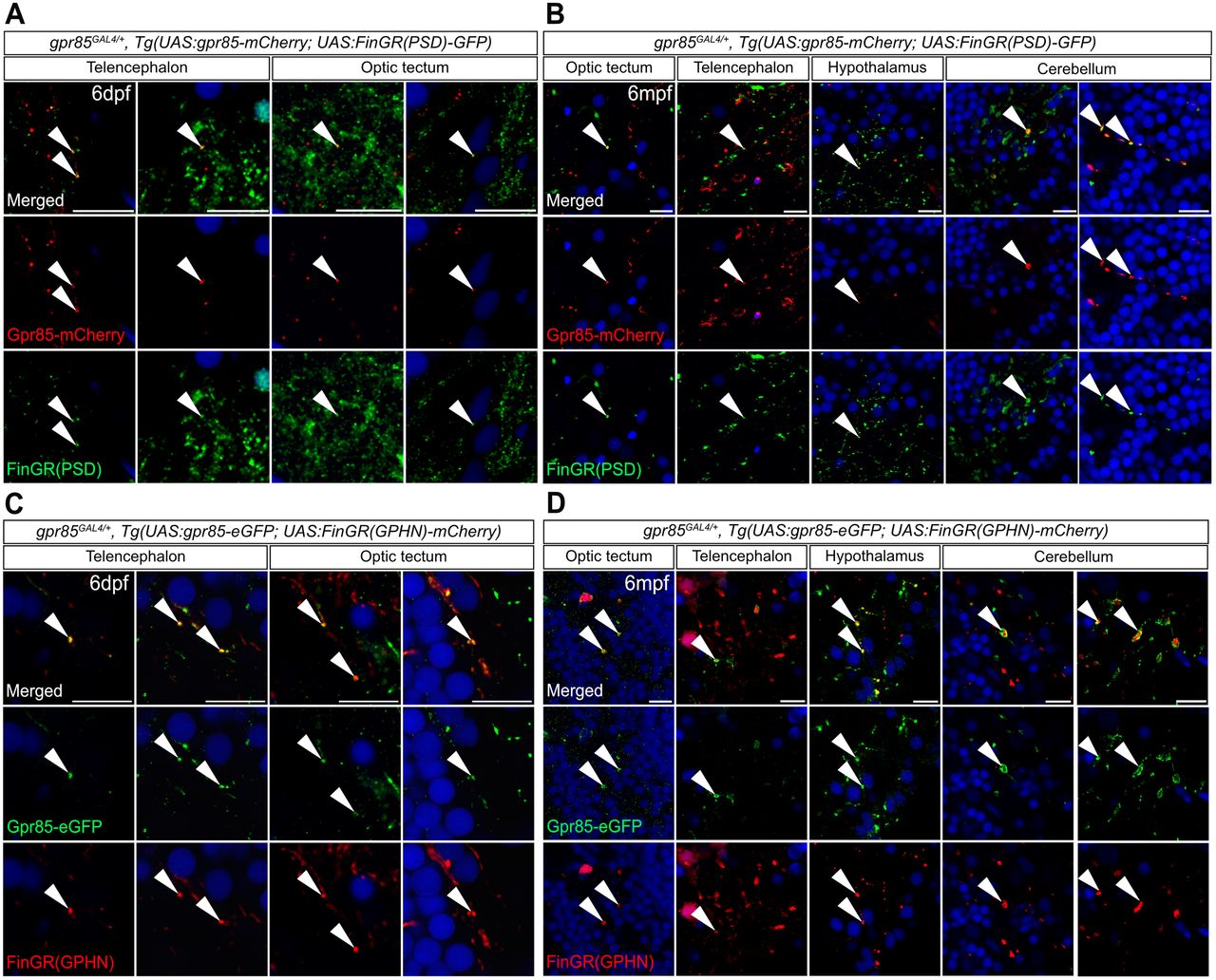
Fig. 4 Gpr85 is enriched at the level of excitatory and inhibitory synaptic inputs of gpr85-expressing neurons throughout the developing and adult brain. A, Confocal images of coronal brain sections from 6 dpf gpr85GAL4/+, Tg(UAS:gpr85-mCherry; UAS:FinGR(PSD)-GFP) larvae stained with anti-GFP (green), anti-mCherry (red), and DAPI (blue). White arrowheads show examples of Gpr85-mCherry signal localized at the level of PSD+ excitatory synapses in the Tel and TeO. Scale bar, 10 µm. B, Confocal images of coronal brain sections from 6 mpf gpr85GAL4/+, Tg(UAS:gpr85-mCherry; UAS:FinGR(PSD)-GFP) adult zebrafish stained with anti-GFP (green), anti-mCherry (red), and DAPI (blue). White arrowheads show examples of Gpr85-mCherry signal localized at the level of PSD+ excitatory synapses in the indicated brain regions. Scale bar, 10 µm. C, Confocal images of coronal brain sections from 6 dpf gpr85GAL4/+, Tg(UAS:gpr85-eGFP; UAS:FinGR(GPHN)-mCherry) larvae stained with anti-GFP (green), anti-mCherry (red), and DAPI (blue). White arrowheads highlight examples of Gpr85-eGFP signal present at the level of the GPHN+ inhibitory synapses in the Tel and TeO. Scale bar, 10 µm. D, Confocal images of coronal brain sections from 6 mpf gpr85GAL4/+, Tg(UAS:gpr85-eGFP; UAS:FinGR(GPHN)-mCherry) adult zebrafish stained with anti-GFP (green), anti-mCherry (red), and DAPI (blue). White arrowheads highlight examples of Gpr85-eGFP signal present at the level of the GPHN+ inhibitory synapses of the indicated brain regions. Scale bar, 10 µm. (for all N = 3) dpf, days postfertilization; mpf, months postfertilization. GPHN, Gephyrin; PSD, postsynaptic density protein 95.