
Fig. 7
- ID
- ZDB-IMAGE-251218-24
- Genes
- Publication
- Le Bleu et al., 2025 - Voltage-gated calcium channels generate blastema Ca2+ fluxes restraining zebrafish fin regenerative outgrowth
- All Figures
- Figures for Le Bleu et al., 2025
|
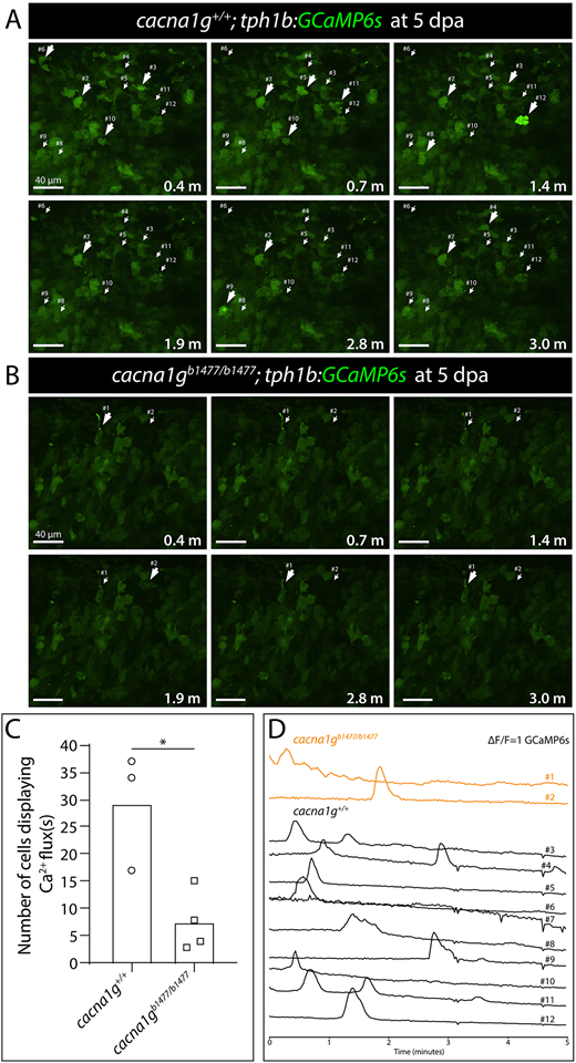
Fig. 7 Cacna1g enables Ca2+ dynamics in fibroblast-derived distal blastema cells of regenerating caudal fins. (A,B) Confocal images from timelapse-imaged regenerating caudal fins of (A) cacna1g+/+ and (B) cacna1gb1477/b1477 fish expressing the tph1b:GCaMP6s reporter. The distal blastema is shown at 5 days post amputation (dpa). Small arrows indicate Ca2+-fluxing cells. Large arrows indicate where Ca2+ levels are elevated. Scale bars: 40 µm. (C) Graph showing the number of Ca2+-fluxing distal fibroblast-lineage blastemal cells in cacna1g+/+ (n=3) and cacna1gb1477/b1477 (n=4) 5 dpa regenerating caudal fins. Each data point represents an individual animal. *P=0.02 by an unpaired Student's t-test. (D) Plots of normalized GCaMP6s intensity traces over a 5-min period for each individual cell marked and numbered in A and B. Orange traces are cacna1gb1477/b1477 cells (n=2) and black traces are cacna1g+/+ cells (n=10).