
Fig. 5
- ID
- ZDB-IMAGE-251218-22
- Publication
- Le Bleu et al., 2025 - Voltage-gated calcium channels generate blastema Ca2+ fluxes restraining zebrafish fin regenerative outgrowth
- All Figures
- Figures for Le Bleu et al., 2025
|
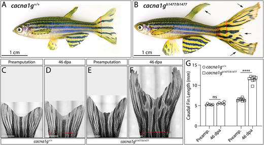
Fig. 5 Long-finned cacna1g loss-of-function mutants link T-type voltage-gated Ca2+ channels to regenerated fin size restoration. (A,B) Bright-field images of representative (A) cacna1g+/+ and (B) cacna1gb1477/b1477 adult zebrafish, including their regenerated caudal fins at 66 days post amputation (dpa). Black dashed lines outline pectoral fins. Green dashed lines indicate where dorsal fins were clipped for genotyping. Red dashed lines indicate caudal fin amputation sites. Arrows indicate regions of exaggerated overgrowth. Scale bars: 1 cm. (C-F) Bright-field stitched caudal fin images of cacna1g+/+ and cacna1gb1477/b1477 fish before amputation and at 46 dpa. Red dashed lines mark amputation positions. D and F are also shown in Fig. S13. Scale bars: 2 mm. (G) Graph showing caudal fin lengths (third fin ray, from the amputation plane to the fin tip) of cacna1g+/+ (circles, n=6) and cacna1gb1477/b1477 (squares, n=8) fish prior to amputation (preamp.) and after regeneration (46 dpa). ****P<0.0001 using paired two-way ANOVA and Tukey's post-hoc tests; ns, not significant (P=0.5).