
|
FIGURE 1
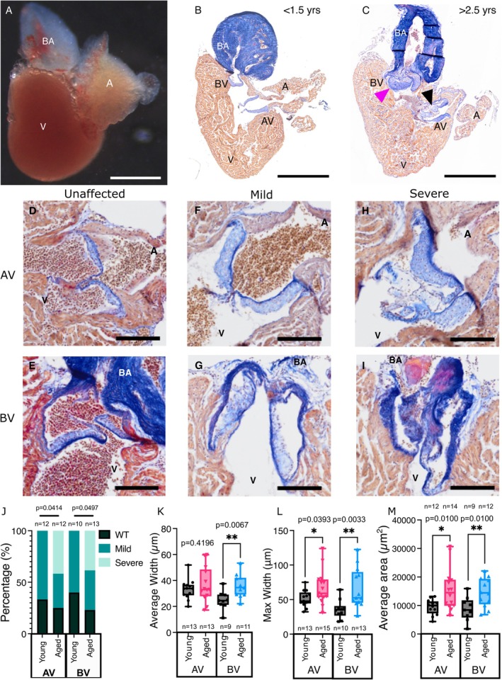
Morphological changes observed in adult zebrafish valves. (A) Image of a dissected heart from a 1‐year‐old zebrafish. (B, C) Overview images of sections of adult zebrafish hearts (< 1.5 years‐B; > 2.5 years‐C) stained with Acid Fuchsin Orange G (AFOG). The atrioventricular (AV) and bulboventricular (BV) valves are indicated. The magenta arrowhead in C indicates thickened leaflets in the BV and the black arrowhead highlights a cystic, nodular region in the AV of an aged zebrafish. (D–I) Higher magnification views of the AV (D, F, H) and BV (E, G, I) indicating examples of phenotypes assigned to the indicated severity for the qualitative scoring shown in J. (J) Qualitive scoring of the AV and BV from young and aged fish. (K–M) Quantification of the average width (K), maximum width (L) and average area (M) of individual leaflets of the AV and BV in young and aged fish. n numbers are indicated on all groups. A, atrium; BA, Bulbus arteriosus; V, ventricle. Statistical analysis: