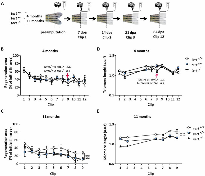
Figure 2
Telomere length is maintained during consecutive amputations in telomerase-deficient zebrafish.
(A) Experimental design of a regeneration assay with 12 consecutive amputations every 7 days and growth monitoring (arrowhead and line), in tert+/+, tert+/−, and tert−/− genotypes, at 4 and 11 months of age. The caudal peduncle is indicated in a blue dotted line. (B) Slopes of the regeneration areas after 12 consecutive clips in the three telomerase genotypes at 4 months of age. n = 3 experiments with 2 biological replicates each. (C) Slopes of the regeneration areas after 9 consecutive clips in the three telomerase genotypes at 11 months age. n = 3 experiments with two biological replicates each. (D) Measurement of telomere length by flow-FISH of regenerative fin tissue cells in all three telomerase genotypes at 4 months of age n = 2 experiments with 1 biological replicate. (E) Measurement of telomere length by Flow-FISH of regenerative fin tissue cells in all three telomerase genotypes at 11 months of age. n = 2 experiments with 1 biological replicate. All data were mean + standard deviation. ***P < 0.001 and ****P < 0.0001 for two-way ANOVA plus Dunnett’s post hoc test, calculated using data at clip number 9. When exact p values are not indicated is because GraphPad Prism software does not show it. Source data are available online for this figure.
Acknowledgments
This image is the copyrighted work of the attributed author or publisher, and
ZFIN has permission only to display this image to its users.
Additional permissions should be obtained from the applicable author or publisher of the image.
Full text @ EMBO Rep.
Your Input Welcome
Thank you for submitting comments. Your input has been emailed to ZFIN curators who may contact you if
additional information is required.
Oops. Something went wrong. Please try again later.