
|
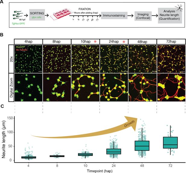
Fig. 3 - Supplemental 1 Zebrafish neuronal primary culture. (A) Schematic summary of the experimental steps in the generation of zebrafish neuronal primary cultures from FACS-sorted HuC:GFP cells. (B) Confocal images of immunofluorescence anti GFP and acetylated tubulin to study zebrafish neuronal outgrowth over time (4, 8, 10, 24, 48, 72 hr after plating [hap]). Top panel: ×20 x confocal images. Bottom panel: digital zoom to show details of neural development progression. Red asterisk shows the two timepoints selected for this study. Scale bars 50 µm. (C) Neurite length manual quantifications of the corresponding confocal images.