IMAGE

Figure 2

ID
ZDB-IMAGE-251127-59
Source
Figures for Dominik et al., 2025
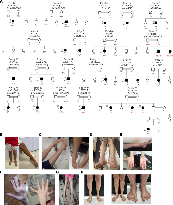
Image
Figure Caption

Figure 2 Genetic and clinical presentation of individuals harboring ARHGAP19 variants.

(A) Pedigrees of affected families showing segregation of the biallelic ARHGAP19 variants identified. Clinical images presenting the spectrum of disease severity in (B) P1 (F1-II:1), (C) P3 (F3-II:1) (D) P10 (F9-II:4) (E) P2 (F2-II:5) (F) P17 (F15-II:3) (G) P20 (F18-II:1) (H) P22 (F21-II:2) and (I) P23 (F21-II:3), with predominant distal lower limb wasting, foot deformity, relatively mild thinning of intrinsic hand muscles and prominent sandal gap (except P17 (F15-II:3) who presented with acute left-hand weakness and still has upper limb–predominant disease).

Acknowledgments
This image is the copyrighted work of the attributed author or publisher, and ZFIN has permission only to display this image to its users. Additional permissions should be obtained from the applicable author or publisher of the image. Full text @ Journal of Clin. Invest.