IMAGE

Figure 5

ID
ZDB-IMAGE-251031-213
Source
Figures for Zeng et al., 2025
Image
Figure Caption

Figure 5

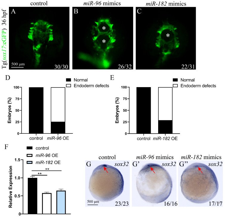
Overexpression of miR-96 and miR-182 leads to defects in endoderm cell convergence. (AC) Fluorescence image of the endoderm of 36 hpf Tg (sox17: eGFP) transgenic zebrafish embryo (ventral view). Control embryos display normal endoderm cell convergence (A); both miR-96 OE (B) and miR-182 OE (C) embryos exhibit endodermal defects with holes in the central (white asterisks). (D,E) Quantification of endodermal defects in miR-96 OE (D) and miR-182 OE (E) embryos. (F) sox32 mRNA expression levels were analyzed by RT-qPCR in control, miR-96 OE, and miR-182 OE embryos at 36 hpf. (GG”) WISH detected the expression of sox32 in control (G), miR-96 OE (G’), and miR-182 OE (G”) embryos at 9 hpf (ventral view; red arrow heads indicate the kupffer’s vesicle). The numbers in the lower right corner indicate the number of embryos. ** p < 0.01.

Acknowledgments
This image is the copyrighted work of the attributed author or publisher, and ZFIN has permission only to display this image to its users. Additional permissions should be obtained from the applicable author or publisher of the image. Full text @ Biomolecules