IMAGE

Figure 3

ID
ZDB-IMAGE-251031-211
Source
Figures for Zeng et al., 2025
Image
Figure Caption

Figure 3

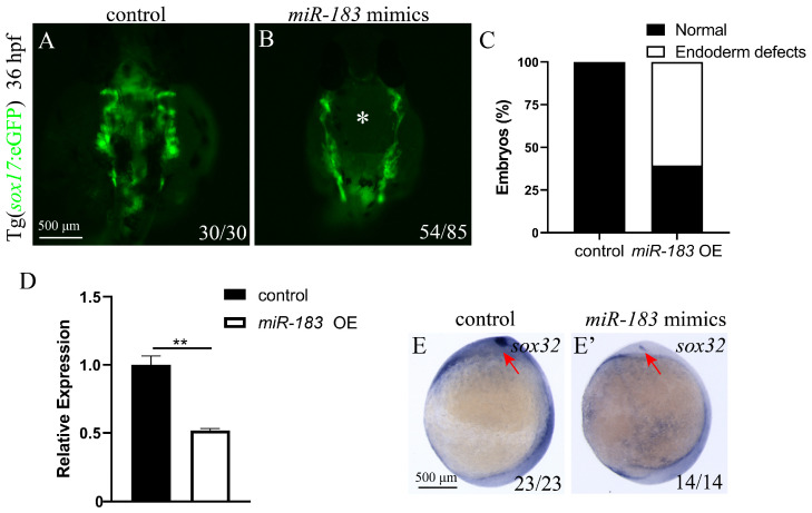
Overexpression of miR-183 disrupts endoderm convergence movement. (A,B) Fluorescence image of the endoderm of transgenic zebrafish embryos with Tg (sox17: GFP) at 36 hpf (ventral view). (A) Control embryos show normal endoderm cell convergence. (B) miR-183 OE embryos exhibit endodermal defects with holes in the central region of the tissue (indicated by white asterisk). Scale bar: 500 μm. (C) Quantification of endodermal convergence defects in miR-183 OE embryos. (D) sox32 mRNA expression levels were analyzed by RT-qPCR in wild-type and miR-183 OE embryos at 36 hpf. (E,E’) WISH detecting sox32 expression in control and miR-183 OE embryos at 9 hpf (ventral view; red arrow heads indicate the kupffer’s vesicle). The numbers in the lower right corner indicate the number of embryos. ** p < 0.01.

Acknowledgments
This image is the copyrighted work of the attributed author or publisher, and ZFIN has permission only to display this image to its users. Additional permissions should be obtained from the applicable author or publisher of the image. Full text @ Biomolecules