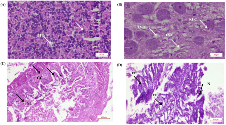
IMAGE

Figure 6

ID
ZDB-IMAGE-251031-192
Source
Figures for Kumari et al., 2025
Image
Figure Caption

Figure 6

Histological analysis of Danio rerio tissues under the effects of CaO-NP-enriched diet: (A) Liver tissue from the negative control group showing normal anatomy. (B) Liver tissue from the T1 group (2.4 mg Cao-NPs/kg feed) showing pyknosis, degenerated hepatocytes, and numerous vacuoles, indicating severe damage and metabolic dysfunction (Note: H—hepatocyte, LVH—liver vacuolation hepatocyte, KLC—Kupffer-like cell; scale bar: 50 µm, magnification: ×400). (C) Intestinal tissue from the negative control group with normal villi and goblet cells. (D) Intestinal tissue from the T1 group (2.4 mg Cao-NPs/kg feed) showing vacuolization of enterocytes, hypertrophy of smooth muscles, pseudo crypts, and muscle layer degeneration, along with damaged goblet cells (Note: GC—goblet cell, FV—fused villi, VE—villar epithelium, N—necrosis, V—villi; scale bar: 100 µm, magnification: ×200).

Acknowledgments
This image is the copyrighted work of the attributed author or publisher, and ZFIN has permission only to display this image to its users. Additional permissions should be obtained from the applicable author or publisher of the image. Full text @ Bioengineering (Basel)