
Image
|
Figure Caption
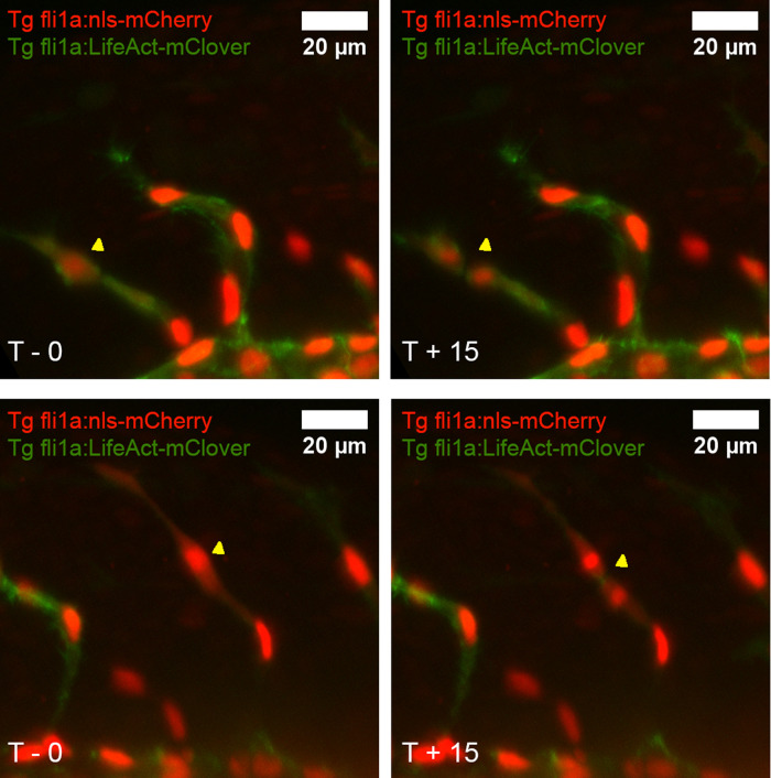
Fig. 4 Endothelial cell proliferation can be directly observed in the intersegmental vessels. The trunk of WT zebrafish was studied from 26 to 30 hpf in 15-minute intervals using a light sheet microscope. Endothelial nuclei were labelled using the transgenic marker fli1a:nls-mCherry (red) whilst endothelial F-actin was labelled using fli1a:lifeAct-mClover (green). EC proliferation was defined as where one nucleus visibly divides into two (examples are shown and dividing cells are labelled with yellow arrows; compare upper left with upper right; compare lower left with lower right).
Acknowledgments
This image is the copyrighted work of the attributed author or publisher, and
ZFIN has permission only to display this image to its users.
Additional permissions should be obtained from the applicable author or publisher of the image.
Full text @ F1000Res