
Image
|
Figure Caption
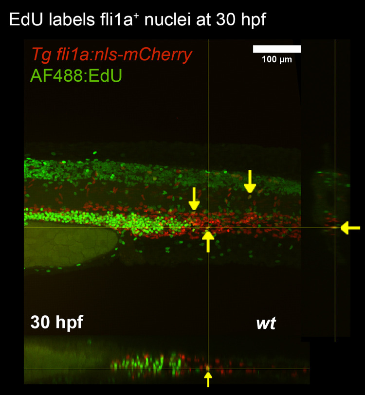
Fig. 3 EdU labelling of zebrafish larvae. Proliferating ECs of Tg ( fli1a:nls-mCherry) embryos were labelled using EdU and visualised using EdU-binding fluorescent azide. Proliferating endothelial nuclei are marked (yellow arrows).Orthagonal views are presented for the X and Y axes.
Acknowledgments
This image is the copyrighted work of the attributed author or publisher, and
ZFIN has permission only to display this image to its users.
Additional permissions should be obtained from the applicable author or publisher of the image.
Full text @ F1000Res