
|
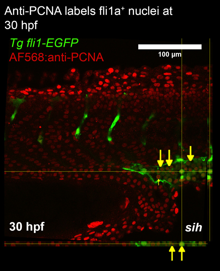
Fig. 2 Whole mount immunostaining of zebrafish larvae for PCNA. Confocal imaging was used to generate a z-stack of fli1a-GFP and PCNA expression in the trunk. Orthogonal views are presented alongside the image. The caudal vein plexus and intersegmental vessels were imaged to determine whether endothelial cell proliferation was detected in these vascular beds. At 30 hpf anti-PCNA was colocalised with fli1a positive cells (labelled with yellow arrows). Since flow modifies EC proliferation, we studied embryos lacking blood flow (tnnt2a morphants; shown) and controls with normal blood flow. PCNA staining did not detect proliferating endothelial cells in either group. The pattern of staining suggests a deficiency of penetration of anti-PCNA antibodies into the centre of the embryo.