
|
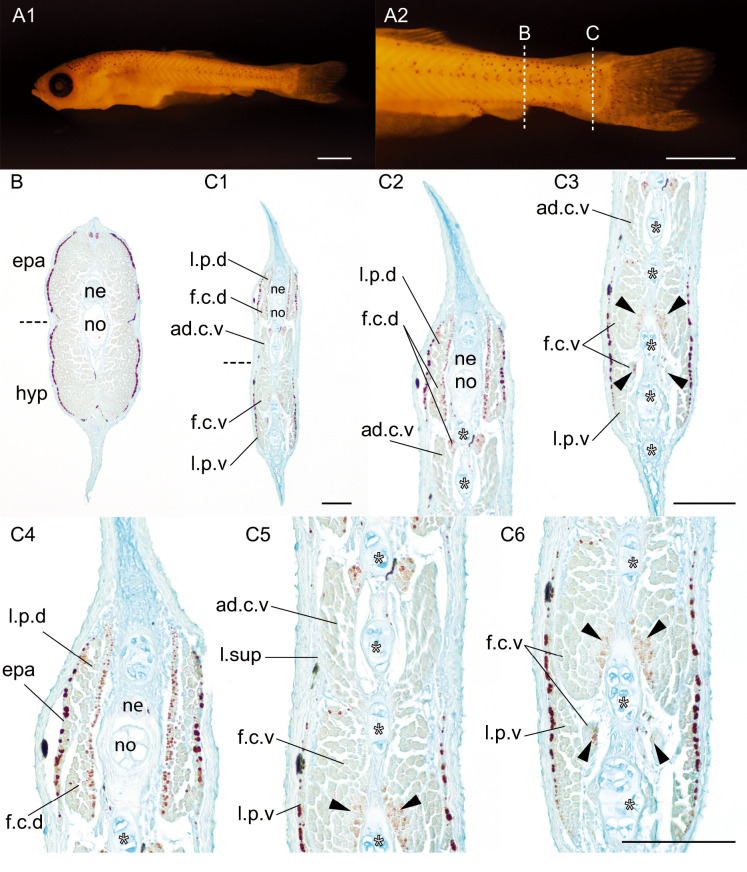
Fig. 3 Transverse section of a wild-type goldfish larva at the late pelvic fin bud stage.(A). Whole lateral view (A1) and magnified view of caudal level (A2) of the goldfish larvae (#2020-0406-01-Bzwj, 7.97 mm [SL], 26 dpf). (B, C). Transverse sections immunostained with the slow muscle fibers specific antibody (F59). (C2, C3). Medium magnification views of dorsal (C2) and ventral (C3) sides at the caudal level sections. (C4-6). High magnification views of dorsal (C4), mid (C5), and ventral regions at the caudal level sections. Sectioned levels are indicated by dashed lines in panel A2. The horizontal myoseptum is indicated by black dashed lines in panels B and C1. White asterisks indicate the ventral caudal skeleton complex including pural, hypural, and hermal spines. Black arrowheads indicate slow muscle fibers in the flexor caudalis ventralis. Abbreviations: ad.c.v, adductor caudalis ventralis; epa, epaxial muscle; f.c.d, flexor caudalis dorsalis; f.c.v, flexor caudalis ventralis; hyp, hypaxial muscle; l.p.d, lateralis profundus dorsalis; l.p.v, lateralis profundus ventralis; l.sup, lateralis superficials; ne, neural tube; no, notochord. Scale bars: A1, A2 = 1 mm; C1, C3, C6 = 100 μm. Panel B and C1, C2 and C3, and panels of the third row have the same magnifications.