
|
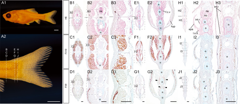
Fig. 2 Transverse section of a wild-type goldfish larva at the pelvic fin ray stage (A). Lateral view of goldfish fixed with Bouin’s fixative (#2021-0517-09-22dpf-B01- ZWJZWJ, 9.95 mm in standard length [SL], 22 dpf). (A). Whole lateral view (A1) and magnified caudal view (A2). (B–J). Transverse sections at the anterior level (B–D), the mid-level (E–G), and the most posterior level (H–J) of the caudal region. The same sections are indicated by the same Roman letters and the magnified views are identified by the plural numeric suffix on the left upper corner of the panels. Dashed boxes are indicated the magnified regions. The upper, middle, and lower panels of the histological sections are conventional histological sections, immunohistochemistry with F310 antibody (the fast muscle fiber), and with F59 antibody (the slow muscle fiber), respectively. White asterisks indicate the ventral caudal skeleton complex including, hypural, parhypural, and hermal spines. Approximate sectioned levels are indicated by dashed lines in panel (A2). Horizontal myoseptum is indicated by black dashed lines in panels B1, B2, and B3. The slow muscle fibers are indicated by black arrowheads in panel G2. The conventional histology, F310, and F59 antibodies-stained sections in the same column are derived from the adjacent sections. Abbreviations: epa, epaxial muscle; f.c.v, flexor caudalis ventralis; hyp, hypaxial muscle; int.r, interradials; l.s.d, lateralis superficialis dorsalis; ne, neural tube; no, notochord. Scale bars: A1, A2 = 1 mm; D1, D2, D3, G1, G1, G2, J1, J3 = 100 μm. Histological sections in the same column have the same magnification.