
Image
|
Figure Caption
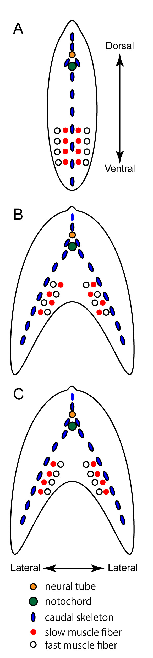
Fig. 15 Schematic representation of the distribution patterns of muscle fibers. Transverse view at the caudal levels of wild-type goldfish and zebrafish (A), that of the mutant showing randomly mixed distribution patterns of the slow muscle fibers (B) (see also Fig. 13), and that of conventional ornamental twin-tail goldfish (C). For simplicity, only the deep ventral muscles are shown.
Acknowledgments
This image is the copyrighted work of the attributed author or publisher, and
ZFIN has permission only to display this image to its users.
Additional permissions should be obtained from the applicable author or publisher of the image.
Full text @ Zool Stud