
|
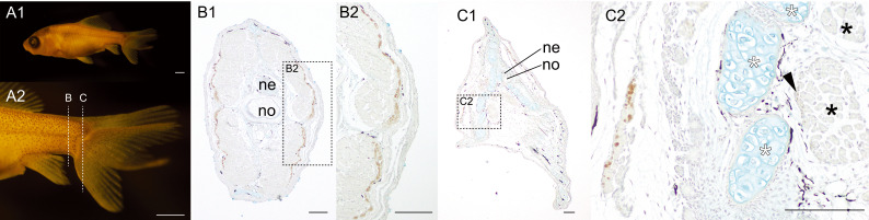
Fig. 14 Distribution pattern of the slow muscle fibers in the 24 twin #02 of the lab goldfish strain.(A, B). Whole body lateral (A1), magnified caudal lateral (A2) of goldfish (#2024-0419-03-#02, 13.09 mm [SL], 23 dpf). (B, C). The different levels of the sections at the caudal region immunostained with the slow muscle specific antibody (F59). The sectioned levels are indicated in the panels of A2. The area of the high magnification views (B2 and C2) are outlined with the dashed boxes in panel B1 and C1, respectively. The medial caudal muscle fibers are indicated by black asterisks in panel C2. White asterisks indicate the ventral caudal skeleton complex including pural, hypural, and hermal spines. Black arrowheads indicate slow muscle fibers in the flexor caudalis ventralis. Abbreviations: ne, neural tube; no, notochord. Scale bars: A, B = 1 mm; C1, D1 = 100 μm; C1, C2, D2 = 10 μm, D3 = 100 μm.