
|
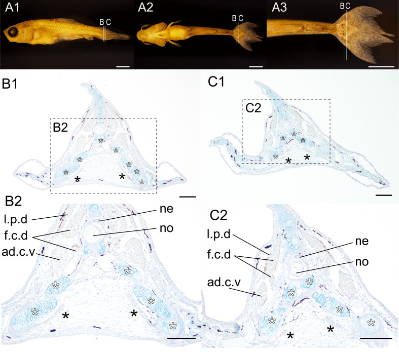
Fig. 11 Distribution patterns of the slow muscle fibers in the 23 wide-twin #01 of the lab goldfish strain. (A). The lateral (A1), the ventral (A2), and the magnified ventral view (A3) of the goldfish (#2023-0417-09-#01, 7.83 mm [SL], 22 dpf). (B–C). The different levels of the sections at the caudal region immunostained with the slow muscle fiber specific antibody (F59). The sectioned levels are indicated in A1. The magnified views are indicated by the dashed boxes with panel labels. White asterisks indicate the ventral caudal skeleton complex including pural, hypural, and hermal spines. The muscular tissues located between the bifurcated caudal fin skeleton are indicated by the black asterisks in panels B1, B2, C1 and C2. Abbreviations: ad.c.v, adductor caudalis ventralis; f.c.d, flexor caudalis dorsalis; l.p.d, lateralis profundus dorsalis; ne, neural tube; no, notochord. Scale bars: A1, A2, A3 = 1 mm; B1, B2, C1, C2 = 100 μm.