
|
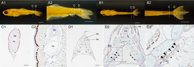
Fig. 10 Distribution pattern of the slow muscle fibers in the 23 narrow twin #08 of the lab goldfish strain.(A, B). Whole body lateral (A1), magnified caudal lateral (A2), whole ventral (B1), and magnified caudal ventral views (B2) of goldfish (#2023-0417-09-#08, 7.16 mm [SL], 22 dpf). (C, D). The different levels of the sections at the caudal region immunostained with the slow muscle specific antibody (F59). The sectioned levels are indicated in the panels of A and B. The first and second columns of section images are low (C1) and high (C2) magnified views at the trunk level. The third, second, and fifth columns of section images are low (D1), mid (D2), and high (D3) magnified views at the caudal level. The area of the high magnification view (D3) is outlined with the dashed box in panel D2. The muscular tissues located between the bifurcated caudal fin skeleton are indicated by black asterisks. White asterisks indicate the ventral caudal skeleton complex including pural, hypural, and hermal spines. Black arrowheads indicate slow muscle fibers in the flexor caudalis ventralis. Abbreviations: ne, neural tube; no, notochord. Scale bars: A, B = 1 mm; C1, D1 = 100 μm; C1, C2, D2 = 10 μm; D3 = 100 μm.