
|
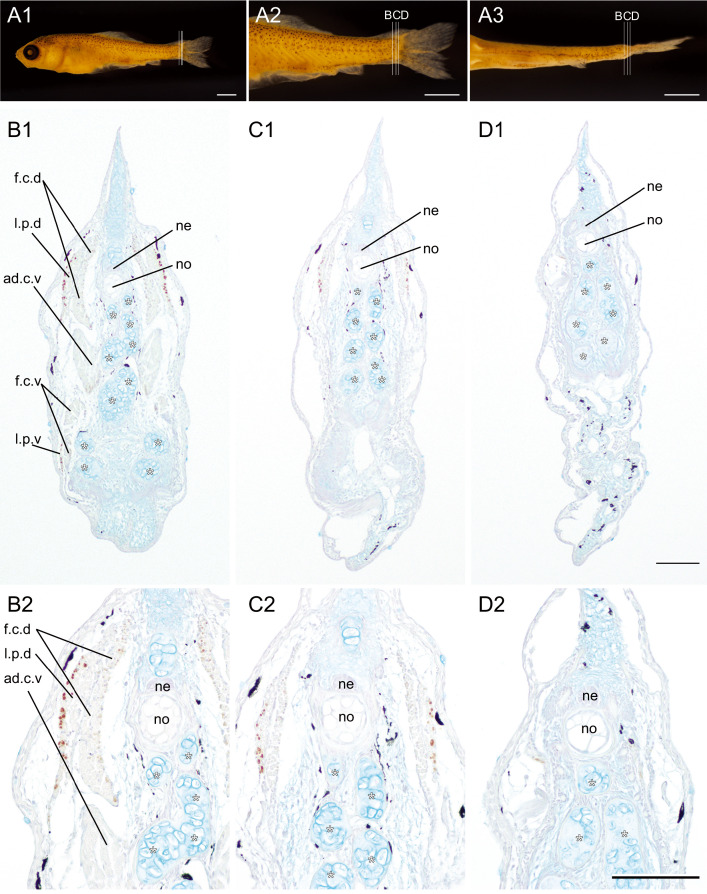
Fig. 9 Distribution pattern of the slow muscle fibers in the 23 single #03 of the lab strain goldfish. (A). Whole body (A1), magnified lateral (A2), and ventral view (A3) of goldfish (#2023-0417-09-#03, 8.59 mm [SL], 22 dpf). The different levels of the sections at the caudal region were immunostained by the slow muscle fiber specific antibody (F59). (B, C, D). The sectioned levels are indicated in A2 and A3. Panels in the second and third rows show the low-magnification (B1, C1, D1) and the high-magnification (B2, C2, D2) images. White asterisks indicate the ventral caudal skeleton complex including pural, hypural, and hermal spines. Abbreviations: ad.c.v, adductor caudalis ventralis; f.c.d, flexor caudalis dorsalis; f.c.v, flexor caudals ventralis; l.p.d, lateralis profundus dorsalis; l.p.v, lateralis profundus ventralis; no, notochord; ne, neural tube. Scale bars: A1, A2, A3 = 1 mm; D1, D2 = 100 μm.