
|
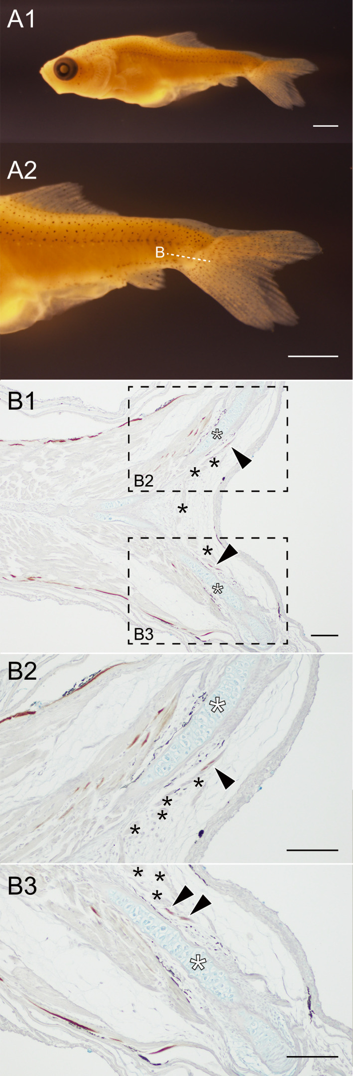
Fig. 7 Horizontal view of the distribution of the slow muscle fibers at the caudal region of Ryukin larva. (A). Whole body (A1) and the magnified caudal region (A2) of Ryukin larva (2022-0502-21-26dof@04, 9.11 mm [SL], 26 dpf). (B). The wide (B1) and magnified views (B2, B3) of horizontal section stained with the slow muscle fiber specific antibody (F59). Magnified areas are indicated by dashed boxes in B1. The muscular tissues located between the bifurcated caudal fin skeleton are indicated by black asterisks. White asterisks indicate the ventral caudal skeleton complex including pural, hypural, and hermal spines. Black arrowheads indicate slow muscle fibers in the flexor caudalis ventralis. Sectioned levels are indicated on panel of A2. Scale bars: A1, A2 = 1 mm; B1, B2, B3 = 100 μm.