
|
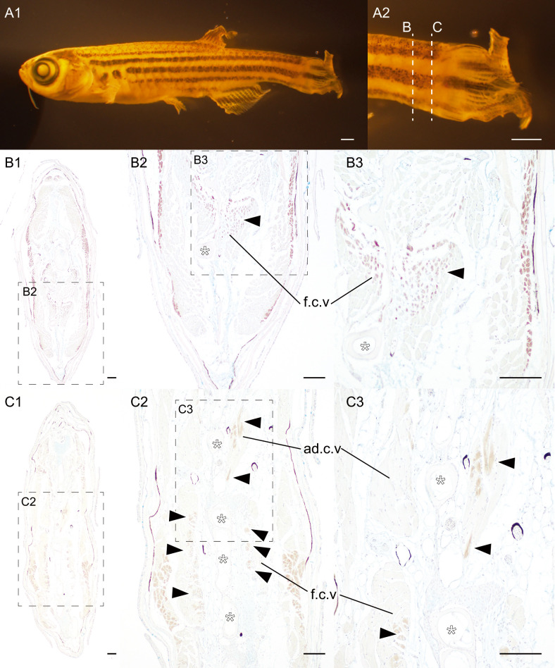
Fig. 5 Slow muscle tissues at the caudal region of zebrafish. (A). The lateral view of zebrafish (2022-0607-ZF-labstrain, 22.0 mm [SL], 582 dpf). (B, C). Transverse sections of immunostained with the slow muscle fiber specific antibody (F59) at caudal fin level. The magnified views of the regions are outlined by the dashed box with panel labels. Panels B and C are derived from two different individuals. Approximate levels of the histological sections are indicated by dashed lines in panel A2. White asterisks indicate the ventral caudal skeleton complex. Black arrowheads indicate slow muscle fibers in flexor caudalis ventralis and adductor caudalis ventralis. Abbreviations: ad.c.v, adductor caudalis ventralis; f.c.v, flexor caudalis ventralis. Scale bars: A = 1 mm; B1–C3 = 100 μm.