
|
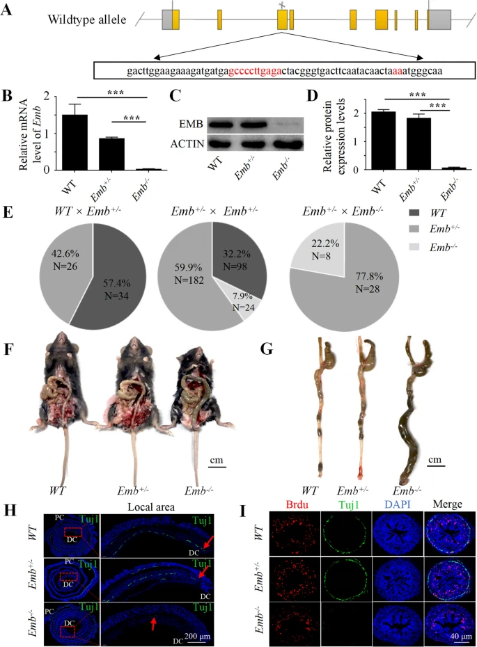
Fig. 2 Loss of Emb causes HSCR-like phenotypes in mice. A, Using CRISPR/Cas9 system, a 13-bp frameshift deletion in exon 3 of Emb was induced. B, Emb mRNA expression is diminished in Emb-/- mice determined by quantitative real-time PCR. C, The EMB protein expression levels in different genotypes were determined by Western blotting, and the expression levels were quantified in (D). E, The percentage of each genotype in three different crossing scenarios. ***, p < 0.001, one-way ANOVA with Bonferroni post-hoc, N = 3. F, Abdominal anatomy of wild type, Emb+/- and Emb-/- mice. G, Isolated colorectal from the distal rectum to the ileocecal part, note the dry stool filled and dilated colon of the Emb-/- mice. H, Immunofluorescence staining of 6-week mouse colons; the entire colon is rolled into a donut-like shape along the longitudinal axis, and then embedded and processed into paraffin sections; the right panels are zoom-ins of the distal colon. Green, Tuj1; blue, DAPI. The red arrows indicate the enteric neurons farthest from the proximal colon, and the distance from the"red arrow"to the distal colon represents the aganglionic segment; PC, proximal colon; DC, distal colon; Quantification results are presented in Additional file 2: Fig.S5 G. I, Immunofluorescence staining of the cross sections of 2-day neonatal mouse colons. Red, Brdu; green, Tuj1; blue, DAPI. Quantification results are presented in Additional file 2: Fig.S5 H