IMAGE

Figure 4

ID
ZDB-IMAGE-251004-90
Source
Figures for Stemerdink et al., 2025
Image
Figure Caption

Figure 4

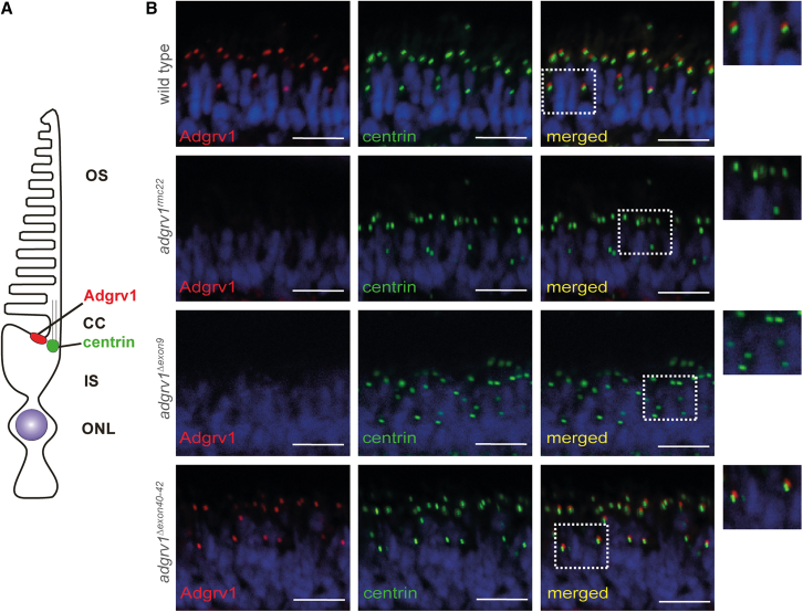
Analysis of Adgrv1 expression in retinal sections of wild-type, adgrv1rmc22, adgrv1Δexon9, and adgrv1Δexon40-42 zebrafish

(A) Schematic representation of a zebrafish photoreceptor cell. OS, outer segment; CC, connecting cilium; IS, inner segment; ONL, outer nuclear layer. (B) Retinal cryosections of wild-type, homozygous adgrv1rmc22, adgrv1Δexon9, and adgrv1Δexon40-42 zebrafish larvae (5 dpf) labeled with antibodies directed against Adgrv1 (red) and centrin (green). Nuclei are counterstained with DAPI (blue). No Adgrv1 signal was detected in adgrv1rmc22 and adgrv1Δexon9 zebrafish retinae. In contrast, Adgrv1 was present and localized adjacent to centrin, a marker of the basal body and connecting cilium, in the retinas of wild-type and adgrv1Δexon40-42 zebrafish larvae (n = 14 larvae per line, from two biological replicates for wild-type, adgrv1rmc22, adgrv1Δexon9, and adgrv1Δexon40-42 larvae). Magnified images are shown at the right side. Scale bars, 10 μm.

Acknowledgments
This image is the copyrighted work of the attributed author or publisher, and ZFIN has permission only to display this image to its users. Additional permissions should be obtained from the applicable author or publisher of the image. Full text @ Mol Ther Nucleic Acids