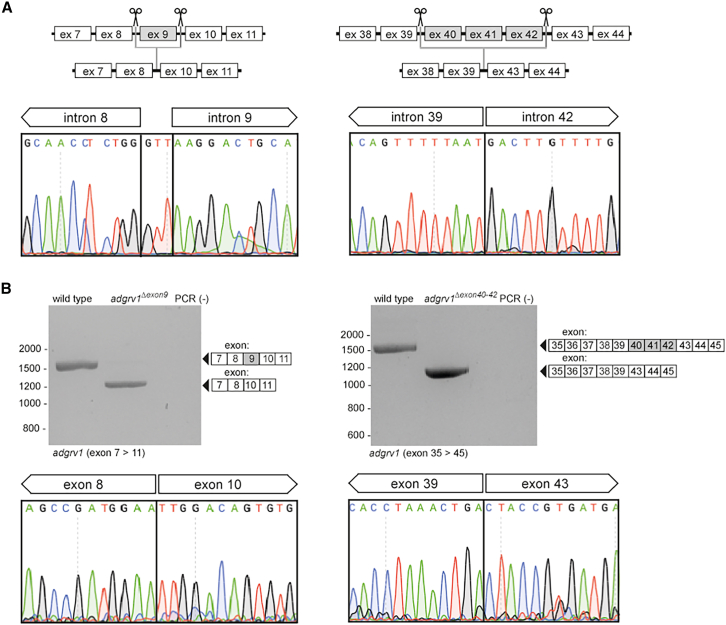
IMAGE

Figure 3

ID
ZDB-IMAGE-251004-89
Source
Figures for Stemerdink et al., 2025
Image
Figure Caption

Figure 3

Design and characterization of the genetically modified adgrv1Δexon9 and adgrv1Δexon40-42 zebrafish lines

(A) Schematic representation illustrating the exon-excision approach. The anticipated excision of the target exons in injected embryos (1 day post-fertilization [dpf]) was confirmed by Sanger sequencing analysis. Excision of the genomic segment encompassing adgrv1 exon 9 led to the incorporation of three additional nucleotides (GTT) at the repair site. (B) RT-PCR results demonstrate the absence of adgrv1 exon 9 in adgrv1Δexon9 larvae, as well as the absence of exons 40–42 in adgrv1Δexon40-42 larvae (5 dpf). Sanger sequencing of RT-PCR amplicons confirmed the successful removal of the target exons from the adgrv1 transcripts. PCR (−): negative PCR control.

Acknowledgments
This image is the copyrighted work of the attributed author or publisher, and ZFIN has permission only to display this image to its users. Additional permissions should be obtained from the applicable author or publisher of the image. Full text @ Mol Ther Nucleic Acids